Review
. 2021 Sep;21(9):597-611.
doi: 10.1038/s41577-021-00519-w. Epub 2021 Mar 26.

Tissue regulatory T cells: regulatory chameleons

Affiliations
Review

Tissue regulatory T cells: regulatory chameleons

Andrés R Muñoz-Rojas et al. Nat Rev Immunol. 2021 Sep.

Abstract

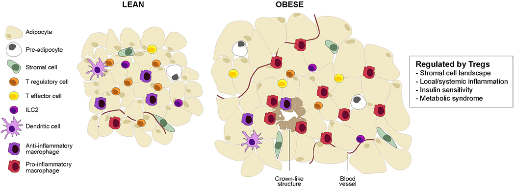
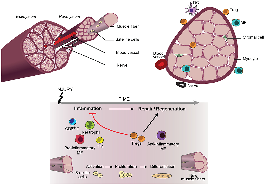
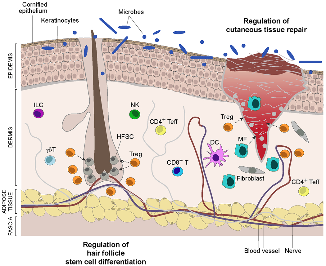
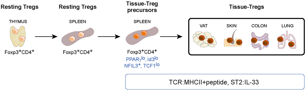
The FOXP3+CD4+ regulatory T (Treg) cells located in non-lymphoid tissues differ in phenotype and function from their lymphoid organ counterparts. Tissue Treg cells have distinct transcriptomes, T cell receptor repertoires and growth and survival factor dependencies that arm them to survive and operate in their home tissue. Their functions extend beyond immune surveillance to tissue homeostasis, including regulation of local and systemic metabolism, promotion of tissue repair and regeneration, and control of the proliferation, differentiation and fate of non-lymphoid cell progenitors. Treg cells in diverse tissues share a common FOXP3+CD4+ precursor located within lymphoid organs. This precursor undergoes definitive specialization once in the home tissue, following a multilayered array of common and tissue-distinct transcriptional programmes. Our deepening knowledge of tissue Treg cell biology will inform ongoing attempts to harness Treg cells for precision immunotherapeutics.


Conflict of interest statement

Competing interests

The authors declare no competing interests. DM is a co-founder of TRex Bio and a consultant for Third Rock Ventures and Pandion Therapeutics.

Figures

Figure 1:
Figure 1:. Visceral adipose tissue biology and regulatory T cells.
Schematic of the epidydimal fat pad of “middle-aged” lean versus obese mice. This visceral adipose tissue (VAT) depot, mostly composed of white-adipose cells, hosts a panoply of resident and recruited innate and adaptive immunocytes. Genetic or diet-induced obesity is associated with chronic, low-grade inflammation that entails secretion of inflammatory cytokines (notably, tumor necrosis factor (TNF), IL-1, IL-6 and interferons) as well as accumulation of inflammatory leukocytes, especially pro-inflammatory macrophages. Inflammation of VAT depots promotes type-2 diabetes and other features of the metabolic syndrome, including insulin resistance, fatty liver disease and heart disease. ILC, innate lymphocyte cell.
Figure 2:
Figure 2:. Skeletal muscle biology and regulatory T cells.
Schematic of hindlimb muscle from a young mouse (top) and its response to acute injury (bottom). Upon injury, mostly quiescent muscle progenitor cells (termed satellite cells) are activated, undergo asymmetric division, and differentiate into post-mitotic precursors, which then fuse to form multi-nucleated myotubes. Myotubes engender new myofibres or fuse to existing ones, followed by a stage of terminal differentiation and growth. This regenerative program is landmarked by a well-defined series of myogenic transcription factor changes. Both innate and adaptive immune-system cells positively or negatively regulate skeletal muscle regeneration. Neutrophils are the earliest of responders, followed by pro-inflammatory macrophagess, CD8+ T cells and T helper 1 (TH1) cells. This initial inflammatory response, which is a requirement for effective regeneration, is followed by a reparative stage dominated by anti-inflammatory macrophages and regulatory T (Treg) cells, both cell types essential for effective regeneration. DC, dendritic cell.
Figure 3:
Figure 3:. Skin biology and regulatory T cells.
Schematic of skin showing a hair follicle and a full-thickness wound. The skin epidermis is composed primarily of keratinocytes that differentiate from basal-layer stem cells while migrating upwards, culminating in a cornified layer that interfaces with the environment. This layer is permeated by hair follicles that cycle through resting (telogen) and growth (anagen) phases, reflecting the activity of hair follicle stem cells (HFSCs). The collagen-rich dermis hosts a variety of stromal-cell types and innate and adaptive immunocyte populations. The deeper adipocyte layer functions as mechanical support and in thermoregulation. Regulatory T (Treg) cells integrate with skin cells to perform a diversity of functions: maintenance of tolerance to local self-antigens; promotion of tolerance to the skin microbiota; prevention of collateral damage during pathogen infections; optimization of cutaneous tissue repair; control of fibrosis; regulation of hair follicle stem cell proliferation, differentiation and fate. They exert their influences by impacting the activities of local immune, stromal and stem cells. DC, dendritic cell.
Figure 4:
Figure 4:. The cellular derivation of tissue regulatory T cells.
FOXP3+CD4+ T cells exit the thymus and enter the circulation, including lymphoid organs such as the spleen. Less than 10% of resting lymphoid-organ regulatory T (Treg) cells undergo an unknown activation event that allows them to escape the circulation and filter through tissues. Treg cells are retained in a tissue that expresses peptide–MHC class II complexes recognized by their T cell receptors (TCRs) and therein undergo definitive specialization. VAT, visceral adipose tissue. NFIL3, nuclear factor interleukin-3-regulated protein 3; TCF1, T cell factor 1; ST2, IL-33 receptor.
Figure 5:
Figure 5:. Analysis of tissue regulatory T cell transcriptomes.
Transcripts differentially expressed between each tissue regulatory T (Treg) cell population and its corresponding lymphoid-tissue control (fold change >2 and FDR <0.10) were calculated using the edgeR package. RNA sequencing datasets for VAT, skin and liver Treg cells are from REF. ; skeletal-muscle Treg-cell data are from REF. ; brain Treg cell-data are from REF. ; and intestinal Treg cell-data are from REF. . a. The compiled matrix of log2 (fold-changes) for each of the tissue-Treg-cell populations was scaled and used to perform principal components analysis (PCA). Principal components 1, 2 and 3 with their proportions of explained variance are plotted. b. UpSet plot depicting inter-tissue intersections for upregulated (top) and downregulated (bottom) transcripts from tissue-Treg cells that had more than 500 differentially expressed genes. The horizontal bar graph indicates the total number of upregulated or downregulated transcripts for each tissue-Treg population. The vertical bar graph shows the number of transcripts corresponding to the particular intersection or set of intersections delineated on the dot matrix. c. Heatmap of log2 (fold-changes) for each tissue-Treg population, separated into pan-tissue, tissue-preferential or tissue-specific gene sets. For each tissue Treg population, the log2 (fold-changes) of transcripts not in the signature (for example, fold-change <2 or FDR >0.10) are set to 0 for that tissue. Example transcripts from the upregulated and downregulated pan-tissue signatures are highlighted on the right. The matrix containing all of the tissue-Treg-cell signatures, whether pan-tissue, tissue-preferential or tissue-specific, can be downloaded at: [https://cbdm.hms.harvard.edu/img/resources/SignaturesAndDatasets/Tissue-Treg%20signatures.xlsx].

Similar articles

Cited by

References

    1. Fontenot JD, Gavin MA, & Rudensky AY Foxp3 programs the development and function of CD4+CD25+ regulatory T cells. Nat. Immunol 4, 330–336 (2003). - PubMed
    1. Hori S, Nomura T, & Sakaguchi S Control of regulatory T cell development by the transcription factor Foxp3. Science 299, 1057–1061 (2003). - PubMed
    1. Khattri R, Cox T, Yasayko SA, & Ramsdell F An essential role for Scurfin in CD4+CD25+ T regulatory cells. Nat. Immunol 4, 337–342 (2003). - PubMed
    1. Ait-Oufella H et al.Natural regulatory T cells control the development of atherosclerosis in mice. Nat. Med 12, 178–180 (2006). - PubMed
    1. Feuerer M et al.Lean, but not obese, fat is enriched for a unique population of regulatory T cells that affect metabolic parameters. Nat Med 15, 930–939 (2009). - PMC - PubMed

Publication types

Substances