

The precuneal cortex: anatomy and seizure semiology
- PMID: 33772513
- PMCID: PMC8525033
- DOI: 10.1684/epd.2021.1257
The precuneal cortex: anatomy and seizure semiology
Abstract
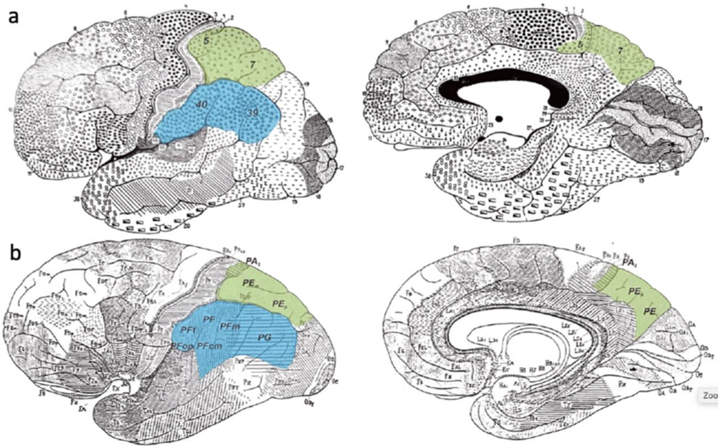
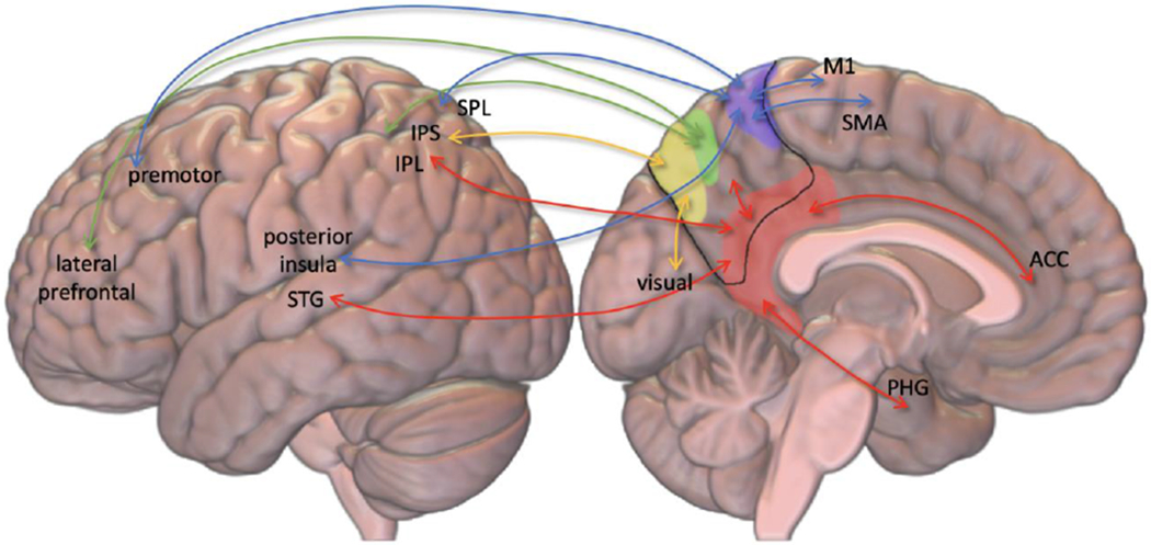
The purpose of this review is to describe the functional anatomy of the precuneal cortex and outline some semiological features of precuneal seizures. The precuneal cortex is a structure that occupies the posterior medial portion of the parietal lobe, and it has broad cortical and subcortical connections. Neuroanatomical tracing, functional imaging, as well as electrical stimulation studies of humans and other primates have elucidated many complex integrative functions of the precuneus including visuo-spatial imagery, sensorimotor functions, and consciousness. Based on the understanding of its functions and connectivity, descriptions of potential seizure semiologies are hypothesized and compared to what is available in the literature. The latter is mostly in the form of case reports or case series. Seizures may involve simple or complex motor or sensory manifestations including abnormal eye movements, visual hallucinations, sensation of motion, or medial temporal-like seizures.
Keywords: SEEG; epilepsy surgery; focal epilepsies; parietal lobe; parietal lobe epilepsy; precuneus.
Conflict of interest statement
Disclosure of Conflicts of Interest
NPP is a member of the Scientific Advisory Board for Dixi Medical USA.
Figures
References
-
- Balestrini S et al. (2015) ‘Multimodal responses induced by cortical stimulation of the parietal lobe: a stereo-electroencephalography study’, Brain: a journal of neurology, 138(Pt 9), pp. 2596–2607. - PubMed
-
- Caspers, Amunts K, and Zilles K, Posterior parietal cortex, The Human Nervous System, pp. 1036–1053, 2012.
Publication types
MeSH terms
Grants and funding
LinkOut - more resources
Full Text Sources
Other Literature Sources
Medical
