. 2021 Mar 27;83(5):50.
doi: 10.1007/s11538-021-00890-8.

Patient-Specific Modelling of Blood Coagulation

Affiliations

Patient-Specific Modelling of Blood Coagulation

N Ratto et al. Bull Math Biol. .

Abstract

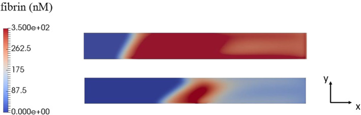
Blood coagulation represents one of the most studied processes in biomedical modelling. However, clinical applications of this modelling remain limited because of the complexity of this process and because of large inter-patient variation of the concentrations of blood factors, kinetic constants and physiological conditions. Determination of some of these patients-specific parameters is experimentally possible, but it would be related to excessive time and material costs impossible in clinical practice. We propose in this work a methodological approach to patient-specific modelling of blood coagulation. It begins with conventional thrombin generation tests allowing the determination of parameters of a reduced kinetic model. Next, this model is used to study spatial distributions of blood factors and blood coagulation in flow, and to evaluate the results of medical treatment of blood coagulation disorders.

Keywords: Blood coagulation; Blood flow; Reaction–diffusion waves; Thrombin generation curves; Treatment.


Figures

Fig. 1
Fig. 1
Schematic representation of the model. It begins with an ODE model for a simplified scheme of coagulation reactions (blue) in order to describe thrombin generation curves and to determine patient-specific parameters (upper left rectangle). The same kinetic model with diffusion is used to describe the propagation of coagulation wave in quiescent plasma (lower left rectangle). A similar model completed by the Navier–Stokes equations and fibrin polymer production describes clot growth in flow (lower right rectangle). Parameters of patient-specific treatment are determined from thrombin generation curves and then used to model clot growth in flow (upper right rectangle) (Color figure online)
Fig. 2
Fig. 2
The main activation reactions of the intrinsic pathway of the coagulation cascade. Thrombin (IIa) catalyses activation of factors V, VIII, XI; factors XIa and IXa catalyse activation of factors IX and X, respectively; factors VIIIa and Va form active complexes with factors IXa and Xa, respectively, and further increase thrombin production. Thrombin accelerates fibrin (F) production from fibrinogen (Fg). Fibrin polymer Fp forms the clot
Fig. 3
Fig. 3
Typical thrombin generation curves for healthy (left) and hemophilia (right) subjects. Solid curves show the fitting of data (crosses) with model (1)–(3). The main parameters characterizing TGCs are shown in the left figure. Reprinted with permission from Ratto et al. (2020a)
Fig. 4
Fig. 4
Normal and hemophilic subjects are separated in the parameter space (k2,k6,k9) characterizing thrombin generation curves in the case of platelet poor plasma. Hemophilic patients are shown by circles, healthy subjects by crosses. Reprinted with permission from Ratto et al. (2020a)
Fig. 5
Fig. 5
Thrombin distributions described by system (4)–(6) in consecutive moments of time (left). Wave speed for healthy subjects (circles) and hemophilia patients (crosses) (right) in μm/s. There are three sub-groups of hemophilia patients: without wave propagation (zero speed), with a slow propagating coagulation wave, with a fast propagating wave
Fig. 6
Fig. 6
Schematic representation of the computational domain corresponding to a part of blood vessel. The damaged vessel wall initiating clot growth is located in the middle of the lower boundary
Fig. 7
Fig. 7
Flow velocity for a final clot shape for hemophilia (upper figure), healthy (middle figure) and thrombotic (lower figure) subjects (shear rate equals 20 1/s). The corresponding fibrin polymer concentration distributions are shown in Fig. 8
Fig. 8
Fig. 8
The concentration of fibrin polymer for a final clot shape for hemophilia (upper figure), healthy (middle figure), and thrombotic (lower figure) subjects. Fibrin clot corresponds to the area with a high concentration of fibrin polymer (red). The corresponding flow velocities are shown in Fig. 7 (Color figure online)
Fig. 9
Fig. 9
Clot height as a function of time for two subjects in each of three groups: thrombotic (two upper curves), healthy (two middle curves), and hemophilic (two lower curves)
Fig. 10
Fig. 10
Dependence of ETP (upper left), lag time (upper right), time to maximum (lower left), and thrombin generation curves on the parameter k9 modelling the action of heparin (its action on k4 is not considered here)
Fig. 11
Fig. 11
Warfarin treatment applied to a virtual patient. The trajectory shows how the characterization of thrombin generation curves (TGC) changes, while the coefficient β (modelling the action of warfarin) is decreased up to 0.67. Treatment does not allow to move the TGC to the zone with healthy subjects (circles)
Fig. 12
Fig. 12
Joint action of heparin and warfarin on three virtual patients. Properly chosen dosage of these drugs brings the parameters of the corresponding TGCs from their original values before treatment (blue circles) to their final values after treatment (green circles). The asterisk indicates the barycenter of the healthy subjects shown by red circles. Treatment is characterized by the coefficients α (heparin) and β (warfarin): 1. α=1.21,β=0.91, 2. α=2.19,β=0.63, 3. α=1.31,β=0.77 (Color figure online)
Fig. 13
Fig. 13
Fibrin polymer distribution for a thrombotic patient (top), and treated thrombotic patient (bottom) for one of the treatment protocols in Fig. 12

References

    1. Ackermann M, et al. Pulmonary vascular endothelialitis, thrombosis, and angiogenesis in Covid-19. N Engl J Med. 2020;383:120–8. doi: 10.1056/NEJMoa2015432. - DOI - PMC - PubMed
    1. Anand M, Rajagopal K, Rajagopal K. A model incorporating some of the mechanical and biochemical factors underlying clot formation and dissolution in flowing blood. J Theor Med. 2003;5(3–4):183–218.
    1. Andreeva AA, Anand M, Lobanov AI, Nikolaev AV, Panteleev MA, Susree M (2018) Mathematical modelling of platelet rich plasma clotting. Pointwise unified model. Russ J Numer Anal Math Modell 33(5):265–276
    1. Beavers G, Joseph D. Boundary conditions at a naturally permeable wall. J Fluid Mech. 1967;30(01):197–207.
    1. Belyaev AV, Panteleev MA, Ataullakhanov FI. Threshold of microvascular occlusion: injury size defines the thrombosis scenario. Biophys J. 2015;109:450–6. - PMC - PubMed

Publication types

LinkOut - more resources