

Hoffa's fat pad thickness: a measurement method with sagittal MRI sequences
- PMID: 33772711
- PMCID: PMC8154775
- DOI: 10.1007/s11547-021-01345-9
Hoffa's fat pad thickness: a measurement method with sagittal MRI sequences
Abstract
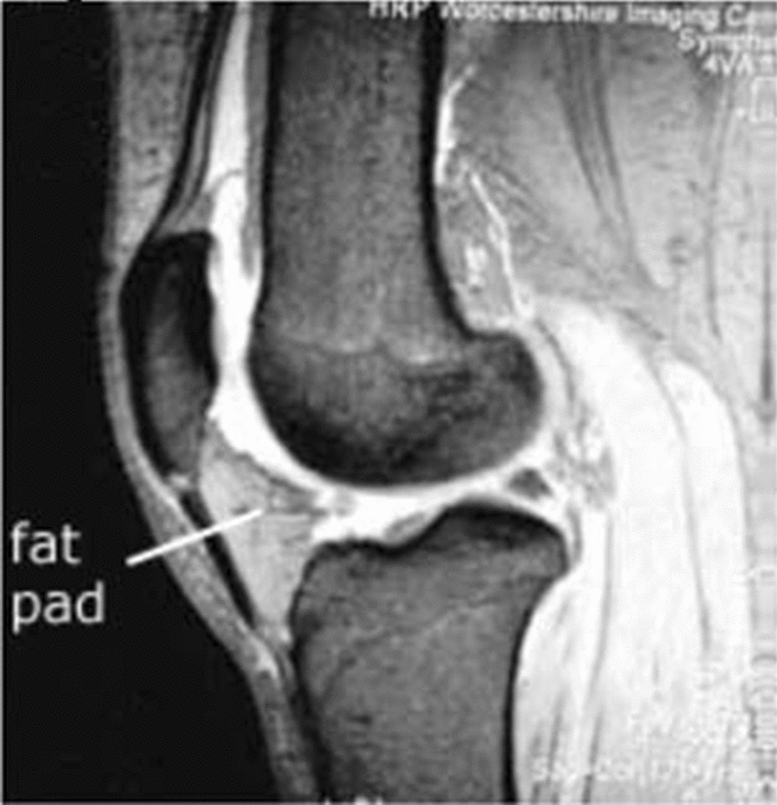
Background: Hoffa's fat pad is a structure located within the fibrous joint capsule of the knee joint, but outside the synovial cavity. It plays an important biomechanical and metabolic role in knee joint, reducing the impact of forces generated by loading and producing cytokines. Changes in its size can induce modifications in the knee homeostasis. However, a great variability exists regarding its measurements. This work aims to evaluate the reliability of a measurement method of Hoffa's fat pad dimensions through MRI.
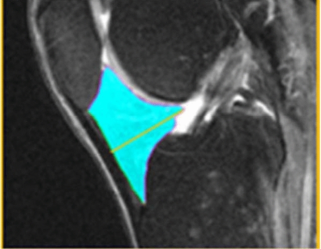
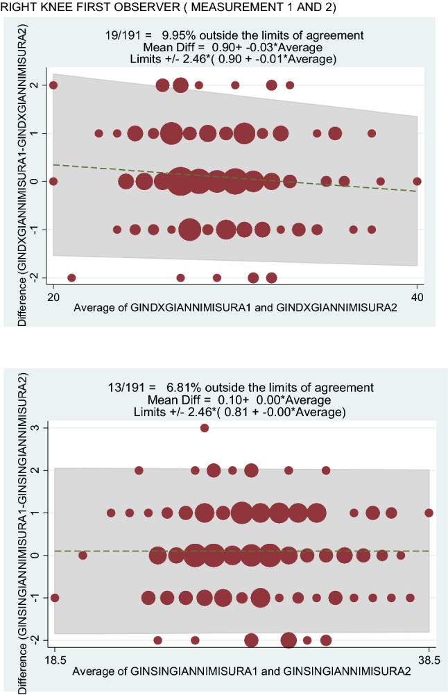
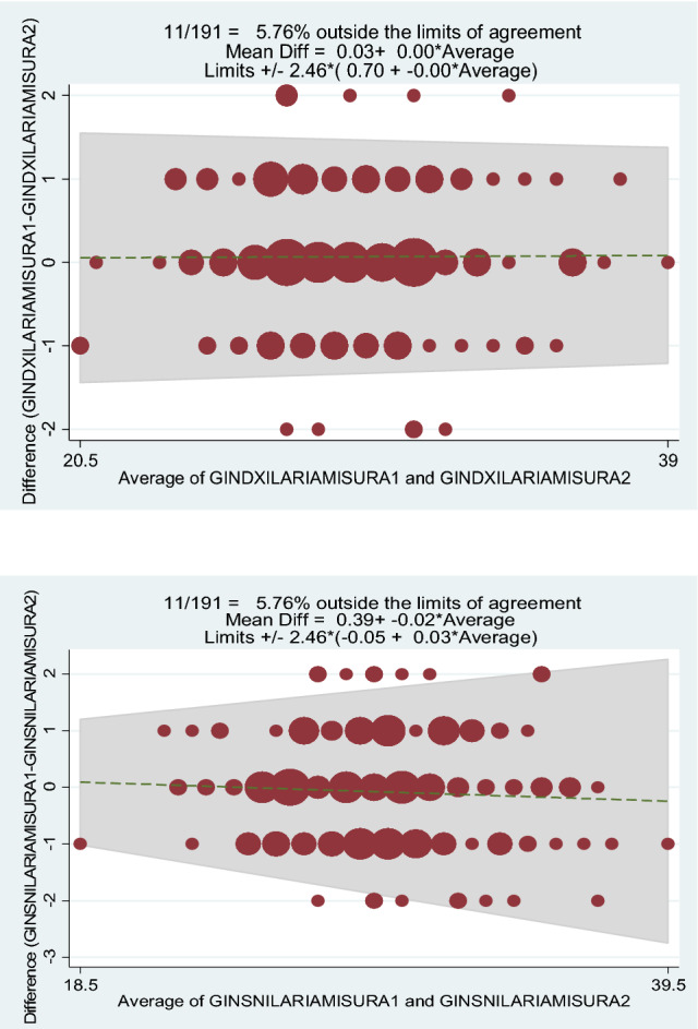
Methods: 3T sagittal IW 2D TSE fat-suppressed MRI sequences, taken from the OAI (Osteoarthritis initiative) database, of 191 male and female patients, aged between 40 and 80 years, were analysed; a manual measurement of the thickness of Hoffa's fat pad of each subject was then performed by two different readers. The interobserver reliability and intraobserver reliability of the measurements were described by coefficient of variation (CV), Pearson correlation and Bland-Altman plots.
Results: All statistical analyses have shown that not significant intra- or interobservers differences were evident (intraobserver CV % for the first observer was 2.17% for the right knee and 2.24% for the left knee, while for the second observer 2.31% for the right knee and 2.24% for the left knee; linear correlation was for the first observer r = 0.96 for the right knee and r = 0.96 for the left knee, while for the second observer r = 0.97 for the right knee and r = 0.96 for the left knee; in addition, the interobserver CV % was 1.25% for the right knee and 1.21% for the left knee and a high interobserver linear correlation was found: r = 0.97 for the right knee and r = 0.96 for the left knee). All results suggest that this manual measurement method of Hoffa's fat pad thickness can be performed with satisfactory intra- and interobserver reliability.
Conclusions: Hoffa's fat pad thickness can be measured, using sagittal MRI images, with this manual method that represents, for his high reliability, an effective means for the study of this anatomical structure.
Keywords: Hoffa’s fat pad; Infrapatellar fat pad; Knee joint; MRI; OAI.
Conflict of interest statement
The authors have no conflicts of interest to declare.
Figures

References
-
- LaPrade RF. The anatomy of the deep infrapatellar bursa of theknee. Am J Sports Med. 1998;26:129e32. - PubMed
-
- Clockaerts S, Bastiaansen-Jenniskens YM, Runhaar J, Van Osch GJ, Van Offel JF, Verhaar JA, et al. The infrapatellar fat pad should be considered as an active osteoarthritic joint tissue: a narrative review. OsteoarthrCartil. 2010;18:876e82. - PubMed
Publication types
MeSH terms
LinkOut - more resources
Full Text Sources
Other Literature Sources
Medical
