. 2021 Mar 1;22(3):935-946.
doi: 10.31557/APJCP.2021.22.3.935.

The Apoptotic Effect of Trichinella spiralis Infection Against Experimentally Induced Hepatocellular Carcinoma

Affiliations

The Apoptotic Effect of Trichinella spiralis Infection Against Experimentally Induced Hepatocellular Carcinoma

Fawzya A Elhasawy et al. Asian Pac J Cancer Prev. .

Abstract

Background: Hepatocellular carcinoma (HCC) is the sixth most common type of cancer. Prognosis of HCC remains unsatisfactory. Therefore, developing new therapeutic modalities is still mandatory. Tumor biotherapy is a novel concept developed as a therapeutic strategy for cancer treatment. There is a similarity between the regulatory mechanism of Trichinella spiralis nurse cell formation and tumor cell apoptosis signal regulation.

Objectives: Induction of apoptosis by T. spiralis can represent a new strategy for tumor treatment.

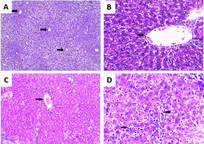
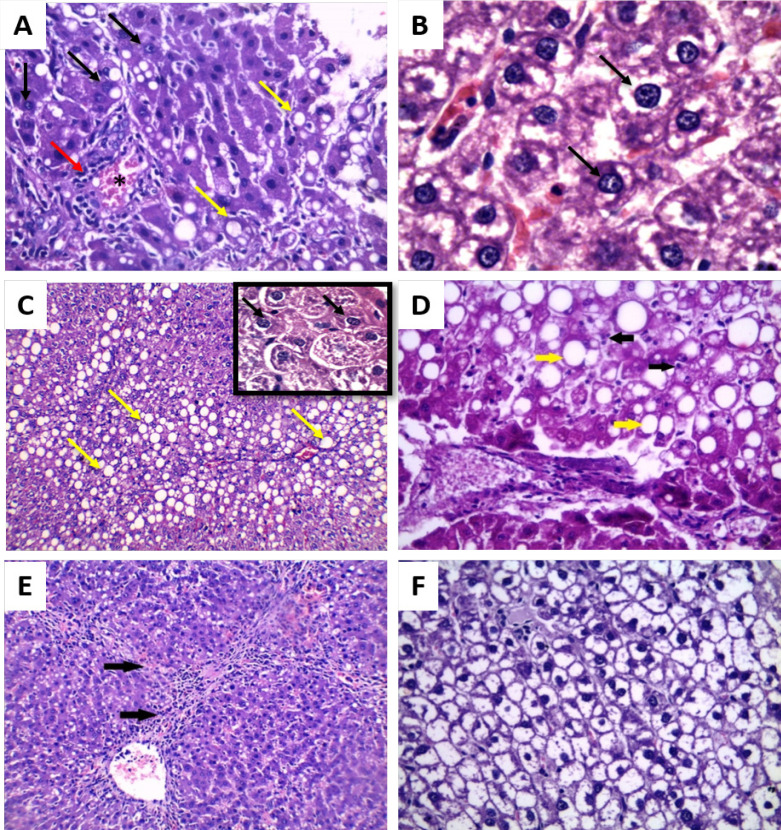
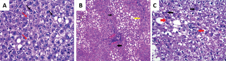
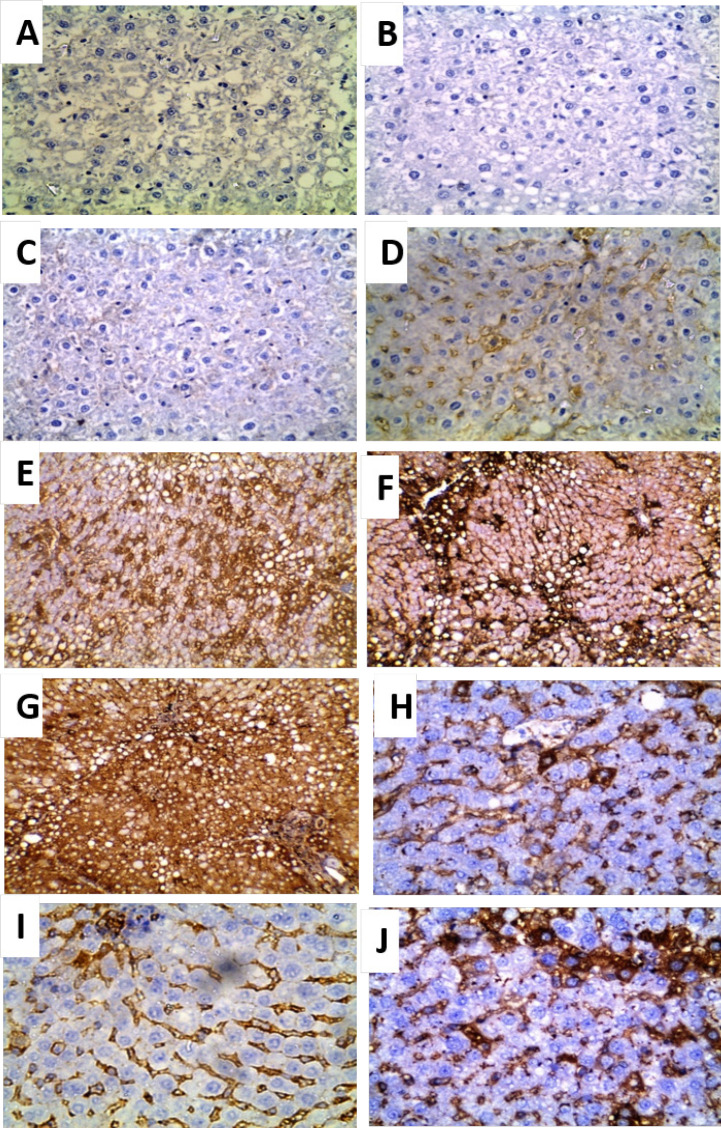
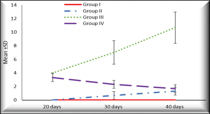
Methods: Experimental animals were divided in four groups; negative control (GI), T. spiralis infected (GII), induced HCC (GIII) and HCC then infected with T. spiralis (GIV). The apoptotic effect of T. spiralis infection was assessed by histopathological and immunohistochemical staining of B-cell lymphoma 2 (Bcl-2).

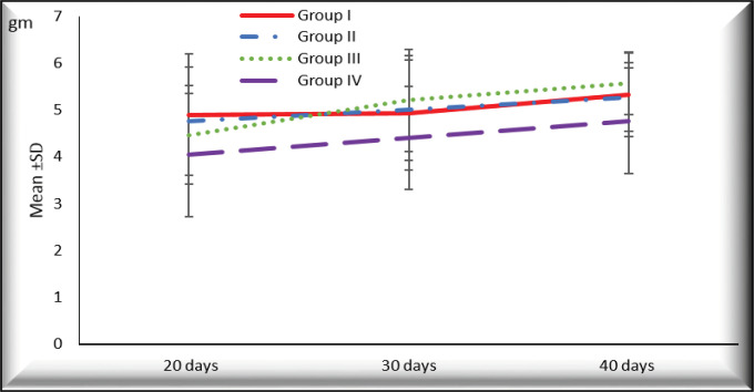
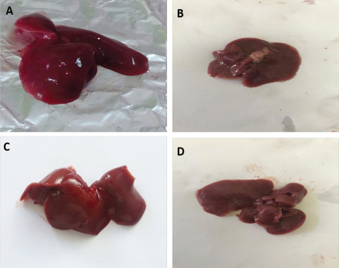
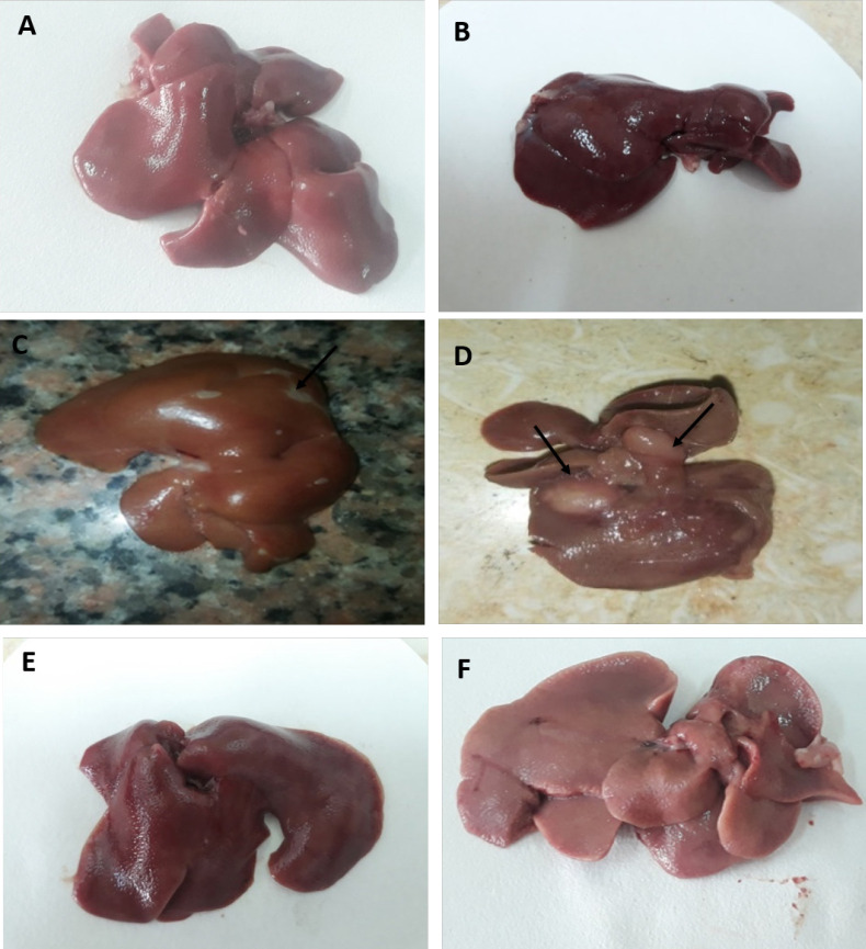
Results: We found higher survival rate of rats and decreased weight of their livers with no nodules in HCC- T. spiralis group as compared to HCC group. Improvement of the dysplastic changes and increased apoptotic bodies which was confirmed by decreased expression of Bcl-2 reported in HCC- T. spiralis group.

Conclusion: Trichinella-induced apoptosis can be a contributing mechanism of the anti-tumor effect of T. spiralis infection. Our results showed a certain level of decreased progression of the tumor in HCC-T. spiralis group as indicated by increased rate of apoptosis and subsequently had a positive impact on the survival of rats.<br />.

Keywords: Apoptosis; Bcl-2; Hepatocellular carcinoma; Trichinella spiralis.


Conflict of interest statement

The authors declare that they have no conflict of interest.

Figures

Figure 1
Figure 1
The Mean Values of Liver Weight of Rats of Different Groups at Different Durations
Figure 2
Figure 2
Photomicrograph of the Liver Showing Its Gross Appearance. A, outer surface; B, inner surface of liver from the negative control rats (group I); C, outer surface; D, inner surface of liver from rats infected with T. spiralis (group II) showing smooth surface, normal color with no macroscopic changes (no hemorrhage, no necrosis, and no masses).
Figure 3
Figure 3
Photomicrograph of the Gross Appearance of Liver of Rats with HCC (group III) (A-D) and HCC- T. spiralis group (group IV) (E-F) showing: (A and B) 20 days from the onset of tumor formation showing dark color of liver and the surface was not smooth, (C and D) outer and inner surfaces of liver 40 days from the onset of tumor formation showing dark color of the liver and the surface was not smooth with some nodules (arrows) and (E-F) outer and inner surfaces of liver showing its gross appearance with slightly darker color and the surface was not smooth
Figure 4
Figure 4
Photomicrograph of Liver Sections (H&E): A- B: negative control rat (group I) showing normal hepatic architecture (arrows showing central veins); A: (×100) and B: ×200) and C-D: T. spiralis infected group (group II); C: showing normal hepatic architecture (arrow showing central veins) (×100) and D: showing apoptotic bodies (arrows) (H&E ×250).
Figure 5
Figure 5
Photomicrograph of Liver Sections of Rats with HCC (group III) (H&E): A: 20 days from the onset of tumor formation showing dysplasia such as increase nucleus size, nucleolus start to appear (black arrow), congestion (asterisk), fatty degeneration (yellow arrow) and infiltration with inflammatory cells mainly lymphocytes (red arrow) (×250), B: 20 days from the onset of tumor formation showing prominent nucleolus, double nuclei (black arrow), clumped chromatin and increased nuclear cytoplasmic ratio (H&E ×400), C: 30 days from the onset of tumor formation showing pleomorphism (multiple prominent eosinophilic nuclei, irregular nuclear membrane (black arrows) and steatosis (yellow arrows) (×250) with insit (×400), D: 30 days from the onset of tumor formation showing few apoptotic bodies (black arrows), steatosis (yellow arrows) (×400), E: 40 days from the onset of tumor formation showing bridging fibrosis (black arrows) and nodules of malignant liver cells (× 250) and F: 40 days from the onset of tumor formation showing clear cell HCC (× 400)
Figure 6
Figure 6
Photomicrograph of Liver Section of HCC- T. spiralis group (group IV) (H&E): A: 20 days from the onset of tumor formation showing apoptotic bodies (red arrows) and dysplastic nuclear changes (black arrows) (× 250), B: 30 days from the onset of tumor formation showing irregular arrangement of hepatocytes, fatty degeneration (yellow arrow) and apoptotic bodies (black arrows) (×100) and C: 40 days from the onset of tumor formation showing apoptosis (red arrows), nuclear dysplasia and pleomorphism (black arrows) (× 400)
Figure 7
Figure 7
Photomicrographs of Immunohistochemical Localization of Bcl-2 in Liver Sections (×400): A: negative control rats (group I) showing negative expression of Bcl-2, B-D: T. spiralis infected rats (group II); B: 20 days p.i. showing negative expression of Bcl-2 (IRS=zero), C: 30 days p.i. showing weak expression of Bcl-2 (IRS=1), D: 40 days p.i. showing moderate expression of Bcl-2 (IRS=2), E- G: HCC group (group III) showing high expression Bcl-2; E: 20 days from the onset of tumor formation (IRS=4), F: 30 days from the onset of tumor (IRS=6) and G: 40 days from the onset of tumor formation (IRS=12) and H-J: HCC- T. spiralis group (group IV) showing weak expression Bcl-2; H: 20 days from the onset of tumor formation (IRS=3 ), I: 30 days from the onset of tumor formation (IRS=2) and J: 40 days from the onset of tumor formation (IRS=1).
Figure 8
Figure 8
The Mean Values of Immunoreactivity Score (IRS) at Different Durations in Different Groups

References

    1. Alenzi FQ, El-Nashar EM, Al-Ghamdi SS, et al. Investigation of Bcl-2 and PCNA in hepatocellular carcinoma: Relation to Chronic HCV. J Egypt Natl Canc Inst. 2010;22:87–94. - PubMed
    1. Babal P, Milcheva R, Petkova S, Janega P, Hurnikova Z. Apoptosis as the adaptation mechanism in survival of Trichinella spiralis in the host. Parasitol Res. 2011;109:997–1002. - PubMed
    1. Badr El-Din NK, Ali DA, Othman R, et al. Chemopreventive role of arabinoxylan rice bran, MGN-3/Biobran, on liver carcinogenesis in rats. Biomed Pharmacother. 2020;126:110064. - PubMed
    1. Berriel E, Russo S, Monin L, et al. Antitumor activity of human hydatid cyst fluid in a murine model of colon cancer. Sci World J. 2013;2013:230176. - PMC - PubMed
    1. Boonmars T, Wu Z, Nagano I, et al. Expression of apoptosis-related factors in muscle infected with Trichinella spiralis. Parasitology. 2004;128:323–32. - PubMed