

doi: 10.1016/j.bpj.2021.03.012.
Epub 2021 Mar 25.
Encapsulation state of messenger RNA inside lipid nanoparticles
Affiliations
- PMID: 33773963
- PMCID: PMC8390897
- DOI: 10.1016/j.bpj.2021.03.012
Item in Clipboard
Encapsulation state of messenger RNA inside lipid nanoparticles
Biophys J.
.
Abstract
Understanding the structure of messenger RNA (mRNA) lipid nanoparticles, and specifically the microenvironment of the mRNA molecules within these entities, is fundamental to advancing their biomedical potential. Here, we show that a permeating cationic dye, thionine, can serve as a cryogenic electron microscopy contrasting agent by binding selectively to encapsulated mRNA without disturbing lipid nanoparticle morphology. Cryo-electron microscopy images identify the mRNA location, revealing that mRNA may exist within solvent-filled cavities or may be substantially lipid associated.
Copyright © 2021 Biophysical Society. Published by Elsevier Inc. All rights reserved.
Figures
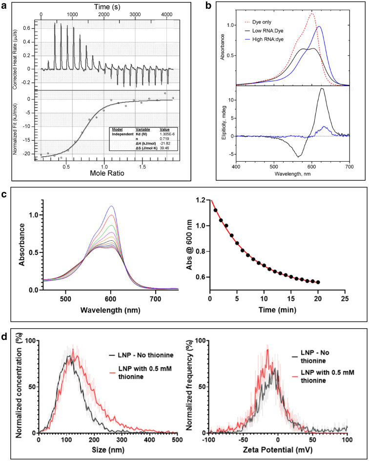
Characterization of dye-binding and permeation. (a) Isothermal titration calorimetry of thionine titrated into mRNA. (Top) Raw data. (Bottom) Integrated heats of each injection versus molar ratio of thionine/nucleotide together with a fit using a one-site binding model. (b) Optical signatures of the thionine-mRNA binding interaction. Visible absorption spectra (top) of thionine at 0.0255 mM recorded in the absence of mRNA and in the presence of mRNA at low (4) and high (160) nucleotide/dye molar ratios (P/D). Corresponding circular dichroism (CD) spectra (bottom) show that at low P/D, the thionine absorption bands are resolved into negative and positive CD bands with extrema at 565 and 628 nm, respectively, whereas at high P/D, the induced CD is weak and characterized by a single positive band at 632 nm. (c) Dye permeation kinetics corresponding to mRNA-LNP added to a thionine solution. (Left) Scanning kinetics showing the spectral change as a function of time with scans taken at 2-min intervals. (Right) Kinetic time course of 600 nm absorbance together with a fit (red) to a first-order process with a rate constant of k = 0.143 min−1. (d) Nanoparticle tracking analysis of mRNA-LNP in the presence and absence of 0.5 mM thionine, showing small or insignificant effects of thionine on particle size and charge. Data represent the mean and error of 3 independent samples. (Left) Size distribution. (Right) ζ potential distribution.
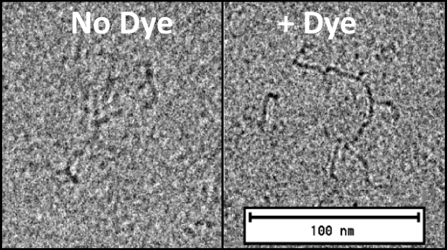
Visualizing free mRNA. Images show contrast enhancement using thionine of a 4000 nucleotide mRNA molecule. In the absence of dye (left), the resolution is comparable to published examples (25), whereas in the presence of 0.1 mM thionine (right), extended strand and molecular branching is more clearly discernible.
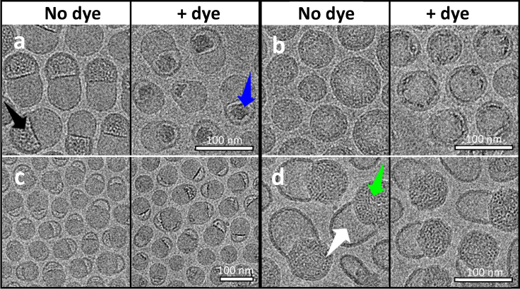
Locating encapsulated mRNA in LNPs by cryo-EM. The effect of thionine on mRNA-LNPs of different morphologies reveals that lipid-dissociated mRNA may reside in bleb compartments (a) or may be more lipid-associated in spherical (b) or less prominently blebbed particles (c). The black arrow in (a) indicates the distinctive mottled mass density of mRNA inside the bleb cavity, which itself is distinguished by a thick, dark periphery. The blue arrow indicates the significant contrast enhancement that occurs when thionine dye is present, thereby identifying mRNA within the bleb. (d) Charge-driven migration of mRNA from the bleb into the body of the LNP. When the mRNA-LNP sample of (a) (no dye) was dialyzed into pH 5 buffer, the images of (d) resulted. The mottled density is now associated with the body of the LNP (green arrow), leaving the bleb cavity devoid of mRNA (white arrow). Addition of thionine to this sample produced no notable contrast change (right), indicating that thionine did not displace lipid.
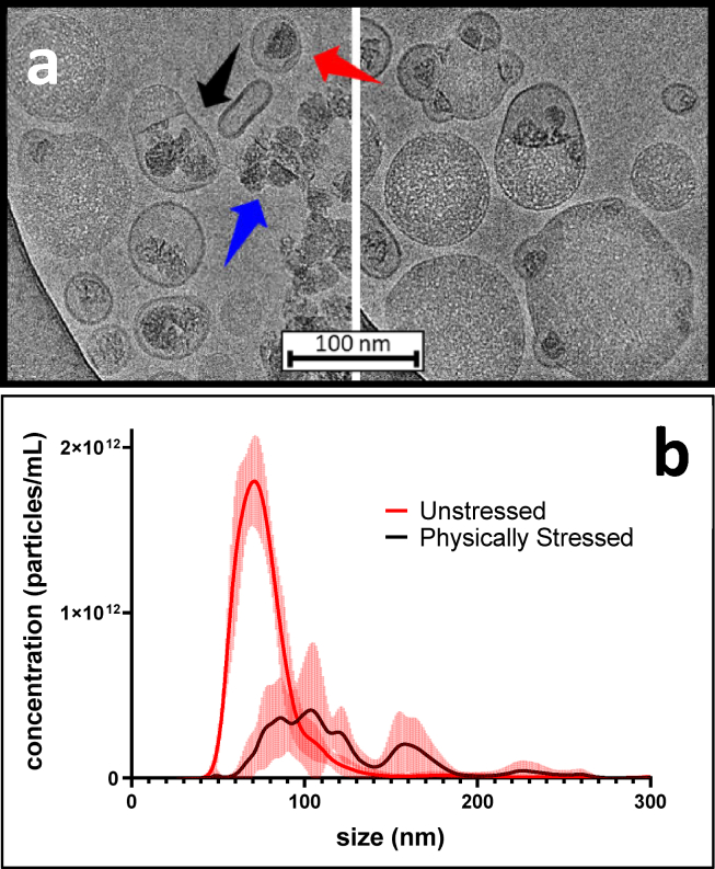
LNP physical degradation pathways. (a) Effects of physical stress on the LNP highlighted by dye. A dye-stained mRNA-LNP sample was subjected to multiple freeze-thaws. The resulting effects of this physical stress are evident, revealing aggregation (right), liberated mRNA (blue arrow), and the formation of liposomal structures containing mRNA (red arrow). The black arrow indicates an LNP that appears ready to burst. This image appears to capture a snapshot of how physical degradation leads to loss of mRNA encapsulation. (b) NTA size distribution profiles corresponding to the stressed and unstressed sample. Mean values with error bars are plotted. A cryo-EM image of the original unstressed LNP sample is provided as Data S2.
References
Publication types
MeSH terms
Substances
LinkOut - more resources
Full Text Sources
Other Literature Sources
