

Prognostication of patients with COVID-19 using artificial intelligence based on chest x-rays and clinical data: a retrospective study
- PMID: 33773969
- PMCID: PMC7990487
- DOI: 10.1016/S2589-7500(21)00039-X
Prognostication of patients with COVID-19 using artificial intelligence based on chest x-rays and clinical data: a retrospective study
Erratum in
-
Correction to Lancet Digit Health 2021; 3: 286-94.Lancet Digit Health. 2021 May;3(5):e283. doi: 10.1016/S2589-7500(21)00060-1. Epub 2021 Mar 31. Lancet Digit Health. 2021. PMID: 33812915 Free PMC article. No abstract available.
Abstract
Background: Chest x-ray is a relatively accessible, inexpensive, fast imaging modality that might be valuable in the prognostication of patients with COVID-19. We aimed to develop and evaluate an artificial intelligence system using chest x-rays and clinical data to predict disease severity and progression in patients with COVID-19.
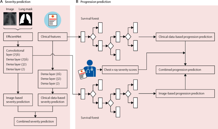
Methods: We did a retrospective study in multiple hospitals in the University of Pennsylvania Health System in Philadelphia, PA, USA, and Brown University affiliated hospitals in Providence, RI, USA. Patients who presented to a hospital in the University of Pennsylvania Health System via the emergency department, with a diagnosis of COVID-19 confirmed by RT-PCR and with an available chest x-ray from their initial presentation or admission, were retrospectively identified and randomly divided into training, validation, and test sets (7:1:2). Using the chest x-rays as input to an EfficientNet deep neural network and clinical data, models were trained to predict the binary outcome of disease severity (ie, critical or non-critical). The deep-learning features extracted from the model and clinical data were used to build time-to-event models to predict the risk of disease progression. The models were externally tested on patients who presented to an independent multicentre institution, Brown University affiliated hospitals, and compared with severity scores provided by radiologists.
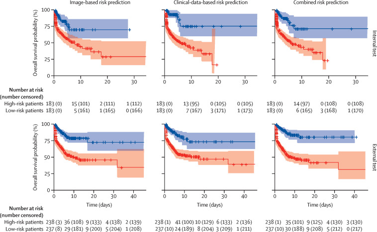
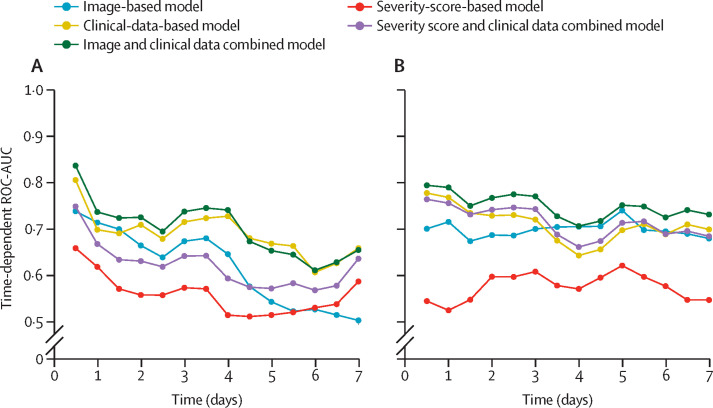
Findings: 1834 patients who presented via the University of Pennsylvania Health System between March 9 and July 20, 2020, were identified and assigned to the model training (n=1285), validation (n=183), or testing (n=366) sets. 475 patients who presented via the Brown University affiliated hospitals between March 1 and July 18, 2020, were identified for external testing of the models. When chest x-rays were added to clinical data for severity prediction, area under the receiver operating characteristic curve (ROC-AUC) increased from 0·821 (95% CI 0·796-0·828) to 0·846 (0·815-0·852; p<0·0001) on internal testing and 0·731 (0·712-0·738) to 0·792 (0·780-0 ·803; p<0·0001) on external testing. When deep-learning features were added to clinical data for progression prediction, the concordance index (C-index) increased from 0·769 (0·755-0·786) to 0·805 (0·800-0·820; p<0·0001) on internal testing and 0·707 (0·695-0·729) to 0·752 (0·739-0·764; p<0·0001) on external testing. The image and clinical data combined model had significantly better prognostic performance than combined severity scores and clinical data on internal testing (C-index 0·805 vs 0·781; p=0·0002) and external testing (C-index 0·752 vs 0·715; p<0·0001).
Interpretation: In patients with COVID-19, artificial intelligence based on chest x-rays had better prognostic performance than clinical data or radiologist-derived severity scores. Using artificial intelligence, chest x-rays can augment clinical data in predicting the risk of progression to critical illness in patients with COVID-19.
Funding: Brown University, Amazon Web Services Diagnostic Development Initiative, Radiological Society of North America, National Cancer Institute and National Institute of Biomedical Imaging and Bioengineering of the National Institutes of Health.
Copyright © 2021 The Author(s). Published by Elsevier Ltd. This is an Open Access article under the CC BY-NC-ND 4.0 license. Published by Elsevier Ltd.. All rights reserved.
Conflict of interest statement
Declaration of interests HXB reports grants from Brown University, Amazon Web Service, Radiological Society of North America, and National Cancer Institute of the National Institute of Health, during the conduct of the study. XF currently works in Carina Medical, a for-profit organisation that develops clinical products, outside of the submitted work. KC reports grants from National Institute of Biomedical Imaging and Bioengineering and National Cancer Institute of the National Institute of Health, during the conduct of the study. YF reports grants from National Institute of Health, during the conduct of the study. All other authors declare no competing interests.
Figures
References
-
- WHO Weekly epidemiological update—2 March 2021. March 2, 2021. https://www.who.int/publications/m/item/weekly-epidemiological-update-2-...
Publication types
MeSH terms
Grants and funding
LinkOut - more resources
Full Text Sources
Other Literature Sources
Medical
