Review
. 2021 Aug 6;433(16):166959.
doi: 10.1016/j.jmb.2021.166959. Epub 2021 Mar 24.

Principles of Alternating Access in Multidrug and Toxin Extrusion (MATE) Transporters

Affiliations
Review

Principles of Alternating Access in Multidrug and Toxin Extrusion (MATE) Transporters

Derek P Claxton et al. J Mol Biol. .

Abstract

The multidrug and toxin extrusion (MATE) transporters catalyze active efflux of a broad range of chemically- and structurally-diverse compounds including antimicrobials and chemotherapeutics, thus contributing to multidrug resistance in pathogenic bacteria and cancers. Multiple methodological approaches have been taken to investigate the structural basis of energy transduction and substrate translocation in MATE transporters. Crystal structures representing members from all three MATE subfamilies have been interpreted within the context of an alternating access mechanism that postulates occupation of distinct structural intermediates in a conformational cycle powered by electrochemical ion gradients. Here we review the structural biology of MATE transporters, integrating the crystallographic models with biophysical and computational studies to define the molecular determinants that shape the transport energy landscape. This holistic analysis highlights both shared and disparate structural and functional features within the MATE family, which underpin an emerging theme of mechanistic diversity within the framework of a conserved structural scaffold.

Keywords: DinF; MATE; NorM; PfMATE; alternating access; antiport; multidrug resistance.


Conflict of interest statement

Declaration of Competing Interest The authors declare that they have no known competing financial interests or personal relationships that could have appeared to influence the work reported in this paper.

Figures

Figure 1.
Figure 1.. MATE phylogenic tree and sequence alignment of subfamily representatives.
(a) Fifty-three sequences derived from the TCDB database [87] were aligned to approximate evolutionary relatedness using the online tool MUSCLE [88]. The phylogram was depicted using the Qiagen CLC Workbench software. Transporters with structural models are highlighted in grey. (b) Sequence alignment of representative MATE transporters, grouped by subfamily, indicating highly conserved residues known to be required for ion and/or substrate binding in the N-lobe (red) and C-lobe (blue). The alignment was performed in Qiagen CLC Workbench using a progressive alignment algorithm.
Figure 2.
Figure 2.. Schematic of alternating access in MATE transporters.
The antiport mechanism requires coordinated interconversion between at least two structural states separated by an energy barrier(s): outward-facing (OF, i and vi) and inward-facing (IF, iii and iv) conformations that expose ligand binding site(s) to opposite sides of the membrane. Within the context of secondary active transport, transduction of potential energy derived from electrochemical ion gradients powers the conformational cycle. Population of other structural intermediates along the transition pathway, including obligatory doubly occluded conformations (ii and v) in which periplasmic and cytoplasmic gates restrict access to binding site(s), play a role in maintaining thermodynamic coupling of ion gradients to transporter mechanics. The steady-state concentration of bound ligand(s) defines the relative occupancy of specific conformations that exist in equilibrium.
Figure 3.
Figure 3.. General topology and fold of MATE transporters.
The representative MATE transporter NorM-Vc (PDB ID 3MKT) displays structural features of the OF conformation. The N- and C- lobes are colored blue and green, respectively. Views from the extracellular (upper right) and intracellular (lower right) sides show 12 transmembrane helices arranged as two symmetric six-helix bundles that constitute the N- and C-lobes and are connected by a long intracellular loop (IL6–7, lower right).
Figure 4.
Figure 4.. Alignment of the N-lobes of NorM-Vc and –Ng reveal structural deviations in the C-lobe and location of ligand binding sites.
NorM-Vc (PDB ID: 3MKU) and NorM-Ng (PDB ID: 4HUK) are depicted as cartoons. The N- and C-lobes for NorM-Vc are colored blue and green, respectively; for NorM-Ng, cyan and yellow, respectively. (a) The ion binding site in the C-lobe of both transporters is formed by conserved residues in the C-lobe (inset) that coordinate sodium congeners Rb+ (purple sphere, 3MKU) and Cs+ (brown sphere, 4HUK). Viewed from the extracellular side of the transporters (b), a 20° tilt of TM7/8 toward the membrane normal is observed in NorM-Ng relative to NorM-Vc. Residues within these helices (b, inset) coordinate the substrate TPP.
Figure 5.
Figure 5.. Comparison of PfMATE with DinF-Bh reveals structural deviations in the C-lobe and distinct substrate binding sites in the N-lobe.
(a) The superimposition reveals TM7/8 displacement in DinF-Bh (PDB ID: 4LZ9, cyan) toward the membrane normal relative to PfMATE (PDB ID: 3VVP, blue). Substrate binding sites are indicated for NFX (dark blue) in PfMATE and R6G (brown) in DinF-Bh. (b) The hydrophobic substrate binding chamber of DinF-Bh stabilizes the conjugated ring system of R6G. (c) A C-lobe fenestration formed by the topological asymmetry of TM7/8 denotes a probable path for substrate extrusion. (d) In addition to participating in NFX binding, Tyr37 of PfMATE engages an H-bond network (dashed lines) involving conserved residues D41 and D184. (e) Mapping of the root mean square deviation (rmsd) between Straight (pH 8.0, PDB ID: 3VVN) and Bent (pH 6.0, PDB ID: 3VVO) conformations of PfMATE onto a ribbon representation of 3VVN indicates the proposed H+-induced structural changes of TM1, 5 and 6. TM1 bending (e, inset), facilitated by the strictly conserved Pro26, collapses the substrate binding cavity.
Figure 6.
Figure 6.. PfMATE intracellular and extracellular gates are mediated by unique molecular interactions.
(a) The IF conformation of PfMATE (PDB ID: 6FHZ) is depicted as a cartoon with the N- and C- lobes colored blue and green, respectively. Helical periodicity of TM1 is disrupted and pivots about Gly30 ~27° in the Y/Z plane and 42° in the X/Z plane. Viewed from the extracellular side (a, upper panel), residues involved in hydrophobic interactions are depicted as sticks while those involved in van der Waals interactions are depicted as spheres (a, inset). Formation of the extracellular gate is concomitant with opening of the central cavity to the intracellular side (a, lower panel). (b) In contrast, the intracellular gate of the OF conformation is mediated by hydrophobic (yellow sticks) and ionic (cyan sticks) interactions (b, middle panel).
Figure 7.
Figure 7.. OF and IF conformational states of MurJ.
(a) MurJ, a putative Lipid II flippase within the MOP superfamily, is composed of 14 TMs arranged as two symmetric six-helix bundles, similar to MATE transporters, but with two ancillary C-terminal TMs that are involved in lipid binding and transport. The N- and C-lobes are colored blue and green, respectively. TMs 13 and 14 of MurJ are depicted as beige ribbons. (b) TM1 from MurJ occupies a number of distinct conformations in the IF state relative to TM1 from PfMATE (blue).
Figure 8.
Figure 8.. Role of conserved residues in the conformational cycle of NorM-Vc.
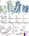
Ligand binding sites in the N- and C-lobe of NorM-Vc (PDB ID: 3MKT) determined from DEER spectroscopy and functional analysis are shown in (a). Critical residues in the C-lobe (left inset) and N-lobe (right inset) are depicted as sticks. The location of spin labels for the reporter 45/269 pair is shown as purple spheres connected by a dashed line. (b) Site directed mutagenesis of Asp36 and Asp371 alter the structural response to ions and substrate, respectively. The distance distributions, P(r), of the reporter pair that retains the functional residues are shaded for each biochemical condition. P(r) for D36N and D371N are shown as solid lines. (c) Loss of DXR binding affinity at low pH is indicated by a right shift in the binding isotherm. Mutation of C-lobe residue Asp371 likewise reduces DXR binding affinity at pH 7.5. (d) Correlation of in vivo DXR resistance activity mediated by NorM-Vc mutants (IC50DXR) with the ion-driven structural transitions quantified by the RMSD between distance distributions. The change in IC50DXR of each mutant corresponds to the color and size of each data point according to the scale.
Figure 9.
Figure 9.. Protons induce population of the IF conformation in PfMATE.
Spin labeled pairs sampling distances between the N- and C-lobes are shown as purple spheres connected with a dashed line and are depicted on the extracellular side (a) and intracellular side (b) of the OF structure (PDB ID: 3VVN). Experimentally-determined distance distributions (solid lines) are plotted with the predicted distance distributions derived from the OF (black, dashed traces) and the IF (red, dashed traces) crystal structures. (a) Measurements from sites on TMs in the C-lobe to sites on TMs in the N-lobe on the extracellular side of the transporter demonstrate H+-dependent decreases in distance between spin labels. (b) In contrast, measurements from sites in the C-lobe to sites in the N-lobe on the intracellular side of the transporter report increases in distance.
Figure 10:
Figure 10:. Protonation of Glu163 facilitates OF-to-IF interconversion in PfMATE.
(a) Glu163 on IL4/5 and Y224 on IL6/7 are depicted as sticks on a cartoon representation of OF PfMATE. (a, bottom panel) Mutation of these residues compromises PfMATE-conferred resistance to toxic concentrations of R6G in a cell based assay. P(r) of E163A and Y224A mutations introduced into extracellular (44/364) and intracellular (95/318) reporter pairs (b, solid lines; upper and lower panels, respectively) is compared with the P(r) of the native construct that retains the wild type residues (shaded distributions) at pH 4 and pH 7.5. The distance components corresponding to the OF and IF conformations on each plot are indicated. E163A and Y224A abrogate the H+-dependent alternating access and destabilize the OF conformation at pH 7.5. (c) A network of polar and charged residues lining the N-lobe could mediate proton translocation from the conserved N-lobe cluster near the periplasmic membrane-water interface to Glu163 on the cytoplasmic side of PfMATE.
Figure 11.
Figure 11.. Putative efflux mechanisms in MATE transporters.
The integration of functional and biophysical studies with crystal structures of MATE transporters highlights mechanistic divergence across subfamilies. The locations of known ion (H+ or Na+) binding sites (orange spheres) are shown relative to the predicted substrate binding sites and proposed permeation pathways (purple region) on cartoon renderings of crystal structure representatives (PDB IDs 5XJJ, 3MKT, 3VVN and 6FHZ, left to right). The structural variation of TM7/8 captured in DinF-Bh (PDB ID 4LZ9, dark blue helices) is mapped onto the structure of PfMATE for comparison. Unlike the DinF subfamily, crystal structures of IF conformations have yet to be determined for eukaryotic and NorM MATE transporters.

References

    1. Du D, van Veen HW, Murakami S, Pos KM & Luisi BF (2015). Structure, mechanism and cooperation of bacterial multidrug transporters. Curr Opin Struct Biol 33, 76–91. - PubMed
    1. Higgins CF (2007). Multiple molecular mechanisms for multidrug resistance transporters. Nature 446, 749–57. - PubMed
    1. Alibert S, N’Gompaza Diarra J, Hernandez J, Stutzmann A, Fouad M, Boyer G & Pages JM (2017). Multidrug efflux pumps and their role in antibiotic and antiseptic resistance: a pharmacodynamic perspective. Expert Opin Drug Metab Toxicol 13, 301–309. - PubMed
    1. Gottesman MM, Fojo T & Bates SE (2002). Multidrug resistance in cancer: role of ATP-dependent transporters. Nat Rev Cancer 2, 48–58. - PubMed
    1. Blair JM, Webber MA, Baylay AJ, Ogbolu DO & Piddock LJ (2014). Molecular mechanisms of antibiotic resistance. Nat Rev Microbiol 13, 42–51. - PubMed

Publication types

Substances

LinkOut - more resources