. 2021 Jan-Jun:296:100586.
doi: 10.1016/j.jbc.2021.100586. Epub 2021 Mar 24.

Rqc1 and other yeast proteins containing highly positively charged sequences are not targets of the RQC complex

Affiliations

Rqc1 and other yeast proteins containing highly positively charged sequences are not targets of the RQC complex

Géssica C Barros et al. J Biol Chem. 2021 Jan-Jun.

Abstract

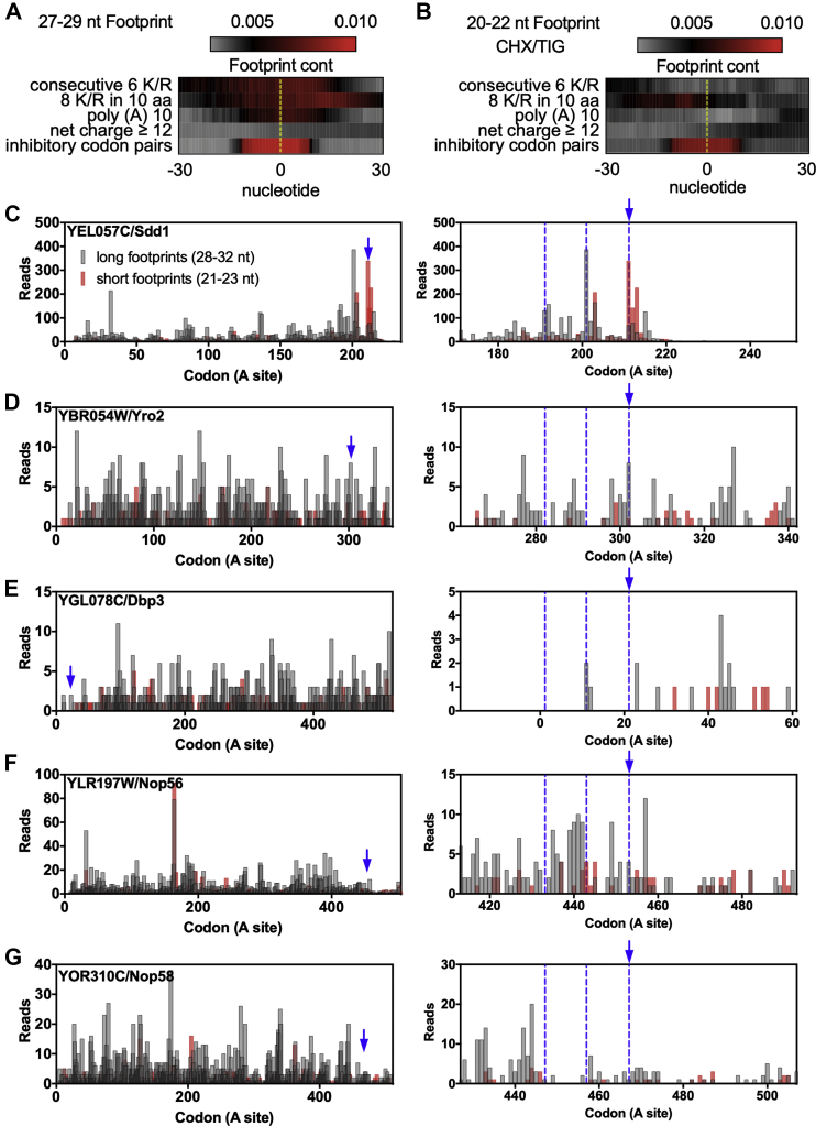
Previous work has suggested that highly positively charged protein segments coded by rare codons or poly (A) stretches induce ribosome stalling and translational arrest through electrostatic interactions with the negatively charged ribosome exit tunnel, leading to inefficient elongation. This arrest leads to the activation of the Ribosome Quality Control (RQC) pathway and results in low expression of these reporter proteins. However, the only endogenous yeast proteins known to activate the RQC are Rqc1, a protein essential for RQC function, and Sdd1, a protein with unknown function, both of which contain polybasic sequences. To explore the generality of this phenomenon, we investigated whether the RQC complex controls the expression of other proteins with polybasic sequences. We showed by ribosome profiling data analysis and western blot that proteins containing polybasic sequences similar to, or even more positively charged than those of Rqc1 and Sdd1, were not targeted by the RQC complex. We also observed that the previously reported Ltn1-dependent regulation of Rqc1 is posttranslational, independent of the RQC activity. Taken together, our results suggest that RQC should not be regarded as a general regulatory pathway for the expression of highly positively charged proteins in yeast.

Keywords: poly (A) tracts; polybasic sequences; protein synthesis; ribosome; ribosome profiling; ribosome quality control; stalled polypeptides; translation; translation control; ubiquitin ligase.


Conflict of interest statement

Conflict of interest The authors declare that they have no conflicts of interest with the contents of this article.

Figures

Figure 1
Figure 1
Bioinformatics screening to identify the polybasic sequences in the yeast proteome.A, Venn diagram showing the genes identified as potential targets of the RQC complex. The list of identified genes is presented in Table S2. B, cumulative frequency distribution of the location of the polybasic sequences of each group. For each gene, the desired attribute was located, and its position was determined in relation to the length of the gene. The frequency of distribution of the genes is depicted in the following categories: (C) consecutive 6 K/R, (D) net charge equal to or higher than +12, and (E) ten or more consecutive adenines. The genes with the highest scores of each category were highlighted.
Figure 2
Figure 2
Translatability scores of genes with polybasic sequences. The different groups of genes with polybasic sequences were compared regarding parameters that, to some extent, reflect the efficiency of translation. A, box plot comparing different parameters from three groups of genes, namely genome, polybasic group, and inhibitory codon pairs (ICP). B, the Kolmogorov–Smirnov test p values are plotted for each comparison. The white rectangle means that a nonsignificant difference was found (p values > 0.05). As a control, genes with one of the 17 inhibitory codon pairs (ICP) were used. C, the values of the translation initiation parameters were normalized (where 0 and 1 represent the lowest and highest initiation rates of the full genome, respectively), and then the 354 identified genes were clustered by the Euclidean distance. The lower panel indicates the polybasic architecture present in each gene (marked in blue). D, translation initiation parameters cluster of the genes with the most prominent polybasic sequences (Fig. 1, CE) and YDR333C/Rqc1 (included for comparison). The raw data, sample size, and p values are presented in Table S4.
Figure 3
Figure 3
Ribosome profiling data of polybasic sequences.A, 27 to 29 and (B) 20 to 22 nucleotide footprint analyses of genes containing polybasic sequences. As control, genes with one of the 17 inhibitory codon pairs (ICP) were used. The dotted yellow line represents the start of the polybasic sequence or of the ICP. C, 28 to 32 and 21 to 23 nucleotide footprint analysis of Sdd1 shows an accumulation of short reads at its polybasic site, indicating ribosome stalling, and accumulation of long reads upstream of the stalling site with a periodicity of roughly ten codons, indicating ribosome collisions, which are highlighted on the right panel. Blue arrow indicates the start of the polybasic sequences, and the blue dotted line indicates the hypothetical position of the collided ribosomes. DG, 28 to 32 and 21 to 23 nucleotide footprint analysis of the proteins with the highest polybasic sequences (YHR131C and YNL143C were omitted because of their low RP coverage). For panels A and B, the full read was used while for panels CG, just the ribosomal A site read was used. The reads were plotted at an approximate position of the ribosomal A site.
Figure 4
Figure 4
Proteins with the greatest polybasic sequences are not targets of the RQC complex in yeast.AF, proteins with the greatest polybasic sequences of yeast were measured by western blot in wild type (wt), lnt1Δ, and asc1Δ strains. For each protein, the primary sequence of their polybasic sequence, their ribosome profiling data, and translation efficiency (TE) are presented. The blue bar in the ribosome profiling data indicates the position of the polybasic sequence, while the blue bar in the TE data shows the TE of the specific gene inside the TE distribution of all genes. The band intensity was quantified and normalized in relation to the housekeeping protein phosphoglycerate kinase (Pgk1). One-way ANOVA with Bonferroni's multiple comparison test was used for statistical analyses.
Figure 5
Figure 5
Rqc1 is not cotranslationally degraded by the RQC complex.A, a TAP-tagged version of Rqc1 was used to measure the protein levels in the wt, lnt1Δ, asc1Δ, rqc2Δ, and hel2Δ strains. The band intensity was quantified and normalized in relation to the housekeeping protein phosphoglycerate kinase (Pgk1). B, qPCR analysis of RQC1-TAP mRNA levels in the wt, lnt1Δ, asc1Δ, rqc2Δ, and hel2Δ strains. The Rqc1 levels were normalized in relation to the housekeeping gene actin (ACT1). The analysis of differential expression was made by relative quantification using 2−ΔΔCt method. One-way ANOVA with Bonferroni's multiple comparison test was used for statistical analyses. C, 28 to 32 and 21 to 23 nucleotide footprint analysis of RQC1 shows neither accumulation of short reads at its polybasic site nor accumulation of long reads upstream of the stalling site (left and right panels, respectively). Blue arrow indicates the start of the polybasic sequences, and the blue dotted line indicates the hypothetical position of the collided ribosomes. The reads were plotted at an approximate position of the ribosomal A site. D, levels of the polybasic reporter GFP-R12-RFP subjected to 10 h cycloheximide (CHX) treatment at increasing concentrations. E, the same experiment as panel D was performed with an Rqc1-TAP strain.

References

    1. Lu J., Deutsch C. Electrostatics in the ribosomal tunnel modulate chain elongation rates. J. Mol. Biol. 2008;384:73–86. - PMC - PubMed
    1. Lu J., Kobertz W.R., Deutsch C. Mapping the electrostatic potential within the ribosomal exit tunnel. J. Mol. Biol. 2007;371:1378–1391. - PubMed
    1. Frischmeyer P.A., van Hoof A., O’Donnell K., Guerrerio A.L., Parker R., Dietz H.C. An mRNA surveillance mechanism that eliminates transcripts lacking termination codons. Science. 2002;295:2258–2261. - PubMed
    1. van Hoof A., Frischmeyer P.A., Dietz H.C., Parker R. Exosome-mediated recognition and degradation of mRNAs lacking a termination codon. Science. 2002;295:2262–2264. - PubMed
    1. Inada T., Aiba H. Translation of aberrant mRNAs lacking a termination codon or with a shortened 3′-UTR is repressed after initiation in yeast. EMBO J. 2005;24:1584–1595. - PMC - PubMed

Publication types

MeSH terms

Substances

LinkOut - more resources