. 2021 Aug;75(2):414-423.
doi: 10.1016/j.jhep.2021.03.016. Epub 2021 Mar 24.

Single-cell atlas of hepatic T cells reveals expansion of liver-resident naive-like CD4+ T cells in primary sclerosing cholangitis

Affiliations

Single-cell atlas of hepatic T cells reveals expansion of liver-resident naive-like CD4+ T cells in primary sclerosing cholangitis

Tobias Poch et al. J Hepatol. 2021 Aug.

Abstract

Background & aims: Little is known about the composition of intrahepatic immune cells and their contribution to the pathogenesis of primary sclerosing cholangitis (PSC). Herein, we aimed to create an atlas of intrahepatic T cells and thereby perform an in-depth characterization of T cells in inflamed human liver.

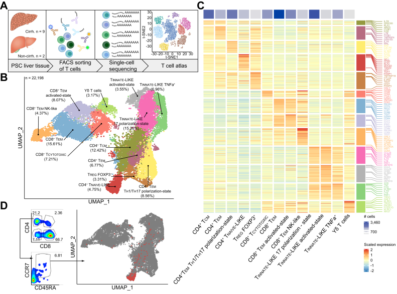
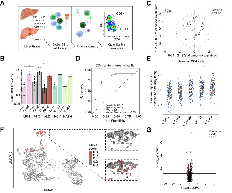
Methods: Different single-cell RNA sequencing methods were combined with in silico analyses on intrahepatic and peripheral T cells from patients with PSC (n = 11) and healthy donors (HDs, n = 4). Multi-parameter flow cytometry and functional in vitro experiments were conducted on samples from patients with PSC (n = 24), controls with other liver diseases and HDs.

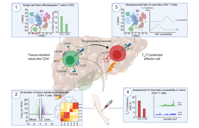
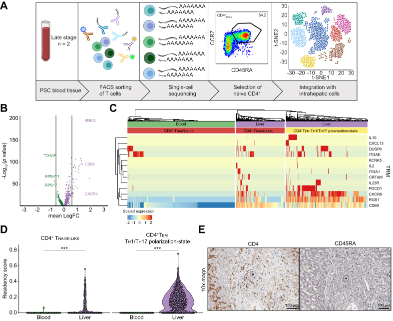
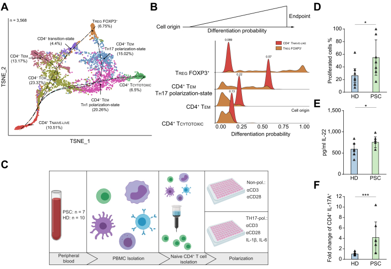
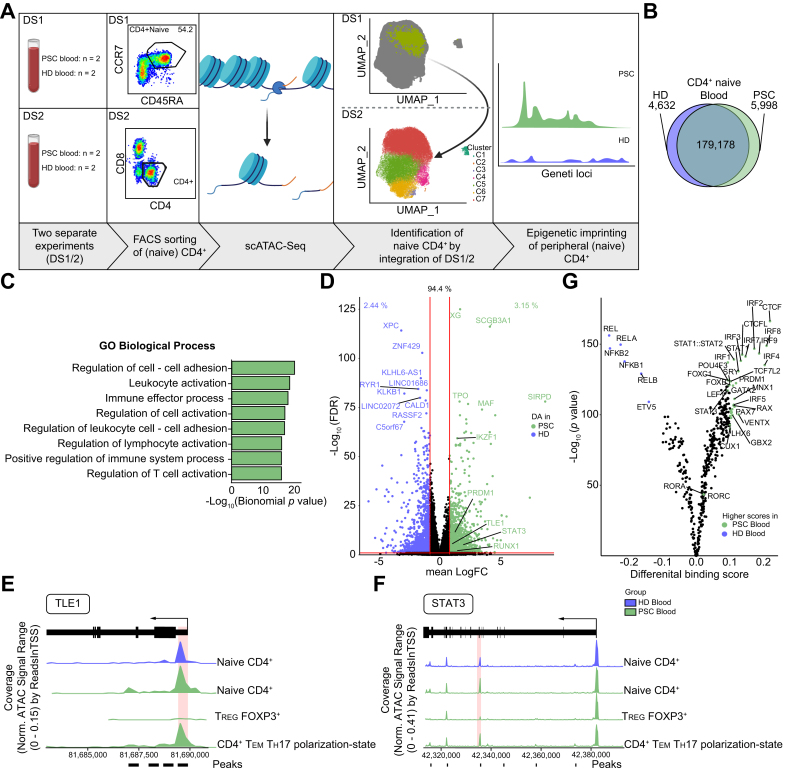
Results: We identified a population of intrahepatic naive-like CD4+ T cells, which was present in all liver diseases tested, but particularly expanded in PSC. This population had a transcriptome and T cell receptor repertoire similar to circulating naive T cells but expressed a set of genes associated with tissue residency. Their periductal location supported the concept of tissue-resident naive-like T cells in livers of patients with PSC. Trajectory inference suggested that these cells had the developmental propensity to acquire a T helper 17 (TH17) polarization state. Functional and chromatin accessibility experiments revealed that circulating naive T cells in patients with PSC were predisposed to polarize towards TH17 cells.

Conclusion: We report the first atlas of intrahepatic T cells in PSC, which led to the identification of a previously unrecognized population of tissue-resident naive-like T cells in the inflamed human liver and to the finding that naive CD4+ T cells in PSC harbour the propensity to develop into TH17 cells.

Lay summary: The composition of intrahepatic immune cells in primary sclerosing cholangitis (PSC) and their contribution to disease pathogenesis is widely unknown. We analysed intrahepatic T cells and identified a previously uncharacterized population of liver-resident CD4+ T cells which are expanded in the livers of patients with PSC compared to healthy liver tissue and other liver diseases. These cells are likely to contribute to the pathogenesis of PSC and could be targeted in novel therapeutic approaches.

Keywords: Atlas; Immune-mediated liver disease; Naive T cells; Primary Sclerosing Cholangitis; Single-cell sequencing; T cells; T(H)17 cells; Tissue residency.


Conflict of interest statement

Conflict of interest The authors declare no conflict of interest. Please refer to the accompanying ICMJE disclosure forms for further details.

Figures

None
Graphical abstract
Fig. 1
Fig. 1
Single-cell atlas of intrahepatic T cells reveals a population of naive-like CD4+ T cells in PSC. (A) Graphical abstract of the workflow on fresh and cryopreserved cells deriving from livers of patients with PSC. (B) Atlas of 22,196 intrahepatic T cells from patients with PSC (n = 11), showing 13 distinct clusters. (C) Cluster-Heatmap highlighting signatures of DEGs for each cluster of the atlas. (D) Hierarchical gated CD4+ T cells, using the ADT for CCR7 and CD45RA, are shown within the atlas, highlighting the naive-like cluster. See also Figs. S1, 6-8 and Table S1. ADT, antibody-derived tags; DEGs, differentially expressed genes; PSC, primary sclerosing cholangitis; TCM, central memory cell; TEM, effector memory cell; TCYTOTOXIC, cytotoxic T lymphocyte; TREG, regulatory T cell; TEM TH1/TH17-state, effector memory cell with a TH1 and TH17 polarization state; UMAP, uniform manifold approximation and projection. Graphical abstract was created with BioRender.com.
Fig. 2
Fig. 2
Intrahepatic naive-like CD4+ T cells are present in inflammatory liver diseases and expanded in patients with PSC. (A) Graphical abstract of the workflow on cryopreserved cells derived from patients with liver diseases of different aetiologies. (B) Frequencies of naive and naive-like CD4+ T cells from blood and liver, respectively, of the same patients determined by classical hierarchical gating for CCR7 and CD45RA. PSC showed the highest frequencies in both liver (PSC vs. ALD, p = 0.0039; PSC vs. LRM, p = 0.0205) and blood (PSC vs. ALD, p = 0.0039). Data in (B) is presented as median (IQR). Statistical significance within blood or liver was assessed by Kruskal-Wallis test (Liver: p = 0.0018; Blood: p = 0.0049). (C) PCA of multi-parameter flow cytometry data of intrahepatic CD4+ T cells from late-stage PSC (grey) and ALD (red). Permutational analysis of variance on Euclidean distance was performed to determine statistical differences between the groups (p = 0.001; R2 = 8.4%). (D) Random forest classifier for intrahepatic CD4+ T cells of PSC and ALD. Discrimination was assessed by AUC, specificity and sensitivity. Statistical significance of the classifier performance was tested by Mason’s and Graham’s non-parametric test. p <0.05 was considered statistically significant. (E) Populations of intrahepatic CD4+ T cells identified to best discriminate between PSC and ALD: identified by iteratively removing the features statistically proven to be less relevant than random samples generated by permutation of the original variable values. (F) UMAP projection of 4,072 intrahepatic T cells of late-stage PSC and ALD. Projection of naive-like DEG core signature, extracted from the atlas, is shown. Cells with matching transcriptomes are highlighted in red. Contribution of underlying disease to the cluster of naive-like CD4+ T cells is shown in the respective colour. (G) Volcano plot of DEG between intrahepatic naive-like CD4+ T cells from late-stage PSC and ALD, indicating similarity of gene expression. Lines indicate cut-off of statistical significance (p <0.05) and logarithmic fold-change of expression (|logFC| >0.7). See also Fig. S2 and Tables S2-3. ALD, alcohol-related liver disease; DEG, differentially expressed gene; LRM, liver resection margin; NASH, non-alcoholic steatohepatitis; PCA, principle component analysis; PSC, primary sclerosing cholangitis; UMAP, uniform manifold approximation and projection. Graphical abstract was created with BioRender.com.
Fig. 3
Fig. 3
Intrahepatic naive-like CD4+ T cells bear a tissue-resident phenotype. (A) Graphical abstract of the workflow on cryopreserved cells deriving from blood and corresponding livers of patients with PSC. (B) Volcano plot of DEG between intrahepatic naive-like CD4+ T cells and bona fide naive CD4+ T cells from the same individuals with PSC showing significant expression of genes accounting for tissue residency. Lines indicate logarithmic fold-change of expression (|logFC|>0.6). (C) Heatmap visualizing gene expression of naive CD4+ T cells coloured by cell origin (green: blood; purple: liver). Expression of a literature-based gene signature for tissue residency is shown. CD4+ TEM TH1/TH17 polarization state from the same dataset was used as positive control. (D) Violin plot quantifying accordance with literature-based residency-score including CD69 (p <0.001 as determined by Wilcoxon signed-rank test). CD4+ TEM TH1/TH17 polarization state from the same dataset was used as positive control (p <0.001 as determined by Wilcoxon signed-rank test). (E) Immunohistochemistry staining of CD45RA and CD4, respectively, in a liver with PSC. Bile ducts are indicated by asterisks and scale bars indicating 100 μm. See also Fig. S3 and Table S4. DEG, differentially expressed gene; PSC, primary sclerosing cholangitis; TEM TH1/TH17-state, effector memory cell with a TH1 and TH17 polarization state; TH1, T helper 1; TH17, T helper 17; TRM, tissue-residency marker. Graphical abstract was created with BioRender.com.
Fig. 4
Fig. 4
Propensity of naive-like CD4+ T cells to develop towards effector cells with a TH17 polarization state in patients with PSC. (A) Diffusion map based t-SNE projection of 3,568 extracted intrahepatic CD4+ T cells from the CITE-Seq dataset of patients with PSC (n = 6, freshly processed samples), showing 8 distinct clusters. Slingshot was applied with naive-like CD4+ T cells defined as starting point. (B) Palantir analysis of the data from (A). Mean probabilities of naive-like CD4+ T cells to differentiate into CD4+ effector memory cells with a TH17 polarization state (CD4+ TEM TH17 polarization-state), CD4+ effector memory (CD4+ TEM), cytotoxic CD4+ (CD4+ TCYTOTOXIC) T cells and regulatory T cells (TREG) were determined. TREG were included as negative control. (C), Graphical abstract of the workflow of in vitro experiments on freshly processed blood samples. (D,E) In vitro culture of blood-deriving naive-like CD4+ T cells under non-polarizing conditions for 7 days (PSC: n = 7; HD: n = 10). (D) Proliferation was determined by detection of CellTrace Violet (Mann-Whitney U; p = 0.0231) and (E) secretion of IL-22 was analysed by multiplex ELISA (Mann-Whitney U; p = 0.0429). (F) In vitro culture of blood-deriving naive-like CD4+ T cells under TH17-polarizing conditions for 12 days (PSC: n = 7; HD: n = 10). Fold change of frequencies of CD4+IL-17A+ cells normalized to the respective controls of each experiment (n = 4) (Mann-Whitney U; p <0.001). Data in (D,E,F) is presented as median (IQR). See also Fig. S4 and Table S5. CITE-Seq, cellular indexing of transcriptomes and epitopes by sequencing; HD, healthy donor; PBMC, peripheral blood mononuclear cell; PSC, primary sclerosing cholangitis; TH17, T helper 17; t-SNE, t-distributed stochastic neighbour embedding. Graphical abstract was created with BioRender.com.
Fig. 5
Fig. 5
Epigenetic predisposition of naive CD4+ T cells in PSC. (A) Graphical abstract of the workflow of scATAC-Seq experiments. Peripheral naive CD4+ and total CD4+ T cells from patients with PSC (n = 4) and HDs (n = 4) were included. Cryopreserved cells were used for these experiments. (B) Venn diagram of shared and differential chromatin accessibility between naive CD4+ T cells from blood of patients with PSC (n = 4) and HDs (n = 4). (C) Volcano plot highlighting mapped regions of shared and significantly different chromatin accessibility between naive CD4+ T cells from blood of patients with PSC (n = 4) and HDs (n = 4). (D) Significantly enriched gene ontology terms regarding biological processes obtained by GREAT analysis of the 5,998 differentially accessible chromatin regions of naive CD4+ T cells between patients with PSC and HDs. (E-F) Coverage plots for selected markers associated with TH17 cell polarization, i.e. TLE1 (E) and STAT3 (F) showing increased chromatin accessibility within naive CD4+ T cells from PSC blood compared to HD blood. Chromatin regions of significantly increased accessibility in PSC are indicated by marks below the coverage plots. Peaks with the lowest FDR/highest Log2FC are highlighted with red boxes. TREG and CD4+ TEM TH17-polarization state were included as a control for visualization. (F) TOBIAS analysis showing differential TFBS accessibility between blood-derived naive CD4+ T cells from patients with PSC (n = 4) and HDs (n = 4). Accessible TFBS of IRFs, STATs, PRDM1 and RORs indicating cellular activation. See also Fig. S5 and Tables S6-7. FDR, false discovery rate; HD, healthy donor; PSC, primary sclerosing cholangitis; TFBS, transcription factor binding site; TH17, T helper 17; TEM TH17 polarization-state, effector memory cells with a TH17 polarization state; TREG, regulatory T cells. Graphical abstract was created with BioRender.com.

References

    1. Karlsen T.H., Folseraas T., Thorburn D., Vesterhus M. Primary sclerosing cholangitis - a comprehensive review. J Hepatol. 2017;67(6):1298–1323. - PubMed
    1. Lazaridis K.N., LaRusso N.F. Primary sclerosing cholangitis. N Engl J Med. 2016;375(12):1161–1170. - PMC - PubMed
    1. Boonstra K., Beuers U., Ponsioen C.Y. Epidemiology of primary sclerosing cholangitis and primary biliary cirrhosis: a systematic review. J Hepatol. 2012;56(5):1181–1188. - PubMed
    1. Liwinski T., Zenouzi R., John C., Ehlken H., Rühlemann M.C., Bang C. Alterations of the bile microbiome in primary sclerosing cholangitis. Gut. 2020;69(4):665–672. - PubMed
    1. Rühlemann M.C., Heinsen F.-A., Zenouzi R., Lieb W., Franke A., Schramm C. Faecal microbiota profiles as diagnostic biomarkers in primary sclerosing cholangitis. Gut. 2017;66(4):753–754. - PubMed

Publication types

MeSH terms