

The duality of PRDM proteins: epigenetic and structural perspectives
- PMID: 33774927
- PMCID: PMC8979648
- DOI: 10.1111/febs.15844
The duality of PRDM proteins: epigenetic and structural perspectives
Abstract
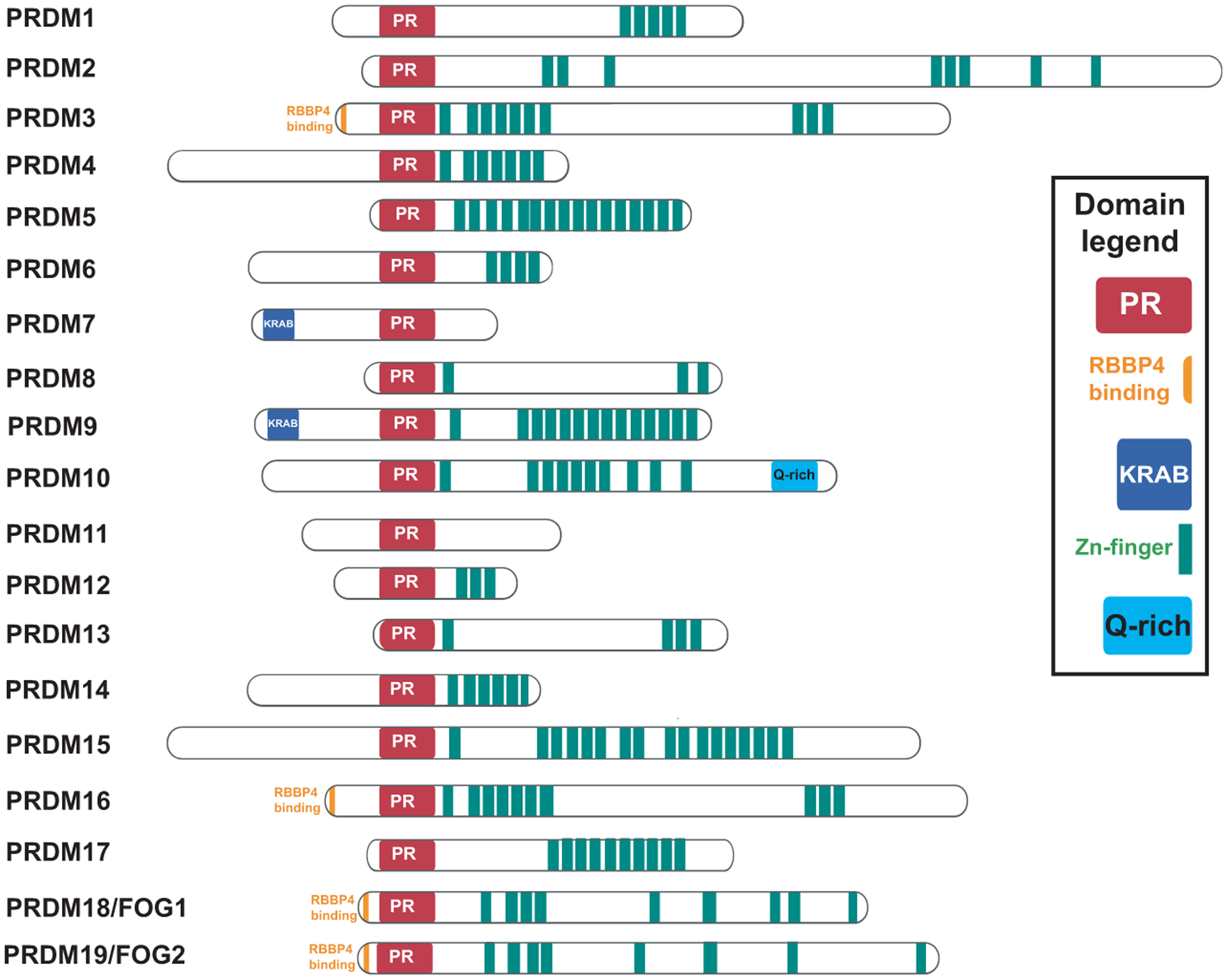
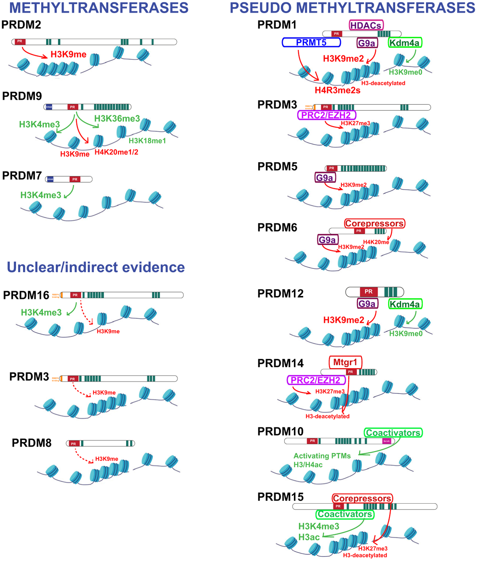
PRDF1 and RIZ1 homology domain containing (PRDMs) are a subfamily of Krüppel-like zinc finger proteins controlling key processes in metazoan development and in cancer. PRDMs exhibit unique dualities: (a) PR domain/ZNF arrays-their structure combines a SET-like domain known as a PR domain, typically found in methyltransferases, with a variable array of C2H2 zinc fingers (ZNF) characteristic of DNA-binding transcription factors; (b) transcriptional activators/repressors-their physiological function is context- and cell-dependent; mechanistically, some PRDMs have a PKMT activity and directly catalyze histone lysine methylation, while others are rather pseudomethyltransferases and act by recruiting transcriptional cofactors; (c) oncogenes/tumor suppressors-their pathological function depends on the specific PRDM isoform expressed during tumorigenesis. This duality is well known as the 'Yin and Yang' of PRDMs and involves a complex regulation of alternative splicing or alternative promoter usage, to generate full-length or PR-deficient isoforms with opposing functions in cancer. In conclusion, once their dualities are fully appreciated, PRDMs represent a promising class of targets in oncology by virtue of their widespread upregulation across multiple tumor types and their somatic dispensability, conferring a broad therapeutic window and limited toxic side effects. The recent discovery of a first-in-class compound able to inhibit PRDM9 activity has paved the way for the identification of further small molecular inhibitors able to counteract PRDM oncogenic activity.
Keywords: yin-yang; PRDM; epigenetic regulation; methyltransferase; pseudomethyltransferase; transcription factor.
© 2021 Federation of European Biochemical Societies.
Conflict of interest statement
Conflict of interest
EG is cofounders and scientific advisors of IMMU-NOA Pte. Ltd. EG has served on advisory board for Lion TCR Pte. Ltd. The rest of the authors declare no competing financial interests.
Figures
Similar articles
-
PRDM proteins: important players in differentiation and disease.Bioessays. 2012 Jan;34(1):50-60. doi: 10.1002/bies.201100107. Epub 2011 Oct 26. Bioessays. 2012. PMID: 22028065 Review.
-
Multifaceted Role of PRDM Proteins in Human Cancer.Int J Mol Sci. 2020 Apr 10;21(7):2648. doi: 10.3390/ijms21072648. Int J Mol Sci. 2020. PMID: 32290321 Free PMC article. Review.
-
The role of PRDMs in cancer: one family, two sides.Curr Opin Genet Dev. 2016 Feb;36:83-91. doi: 10.1016/j.gde.2016.03.009. Epub 2016 May 7. Curr Opin Genet Dev. 2016. PMID: 27153352 Review.
-
PR/SET Domain Family and Cancer: Novel Insights from the Cancer Genome Atlas.Int J Mol Sci. 2018 Oct 19;19(10):3250. doi: 10.3390/ijms19103250. Int J Mol Sci. 2018. PMID: 30347759 Free PMC article.
-
Multifunctional Involvement of a C2H2 Zinc Finger Protein (PbZfp) in Malaria Transmission, Histone Modification, and Susceptibility to DNA Damage Response.mBio. 2017 Aug 29;8(4):e01298-17. doi: 10.1128/mBio.01298-17. mBio. 2017. PMID: 28851851 Free PMC article.
Cited by
-
Updated understanding of the protein-DNA recognition code used by C2H2 zinc finger proteins.Curr Opin Struct Biol. 2024 Aug;87:102836. doi: 10.1016/j.sbi.2024.102836. Epub 2024 May 15. Curr Opin Struct Biol. 2024. PMID: 38754172 Free PMC article. Review.
-
Chromatin Regulator-Related Gene Signature for Predicting Prognosis and Immunotherapy Efficacy in Breast Cancer.J Oncol. 2023 Jan 30;2023:2736932. doi: 10.1155/2023/2736932. eCollection 2023. J Oncol. 2023. PMID: 36755810 Free PMC article.
-
Maternal PRDM10 activates essential genes for oocyte-to-embryo transition.Nat Commun. 2025 Feb 24;16(1):1939. doi: 10.1038/s41467-025-56991-8. Nat Commun. 2025. PMID: 39994175 Free PMC article.
-
Unveiling the Role of Histone Methyltransferases in Psoriasis Pathogenesis: Insights from Transcriptomic Analysis.Int J Mol Sci. 2025 Jun 30;26(13):6329. doi: 10.3390/ijms26136329. Int J Mol Sci. 2025. PMID: 40650108 Free PMC article.
-
PRDM10 directs FLCN expression in a novel disorder overlapping with Birt-Hogg-Dubé syndrome and familial lipomatosis.Hum Mol Genet. 2023 Mar 20;32(7):1223-1235. doi: 10.1093/hmg/ddac288. Hum Mol Genet. 2023. PMID: 36440963 Free PMC article.
References
Publication types
MeSH terms
Substances
Grants and funding
LinkOut - more resources
Full Text Sources
Other Literature Sources
Research Materials
