Review
. 2021 Jul:21:313-330.
doi: 10.1016/j.jtos.2021.03.003. Epub 2021 Mar 26.

Membrane-associated mucins of the human ocular surface in health and disease

Affiliations
Review

Membrane-associated mucins of the human ocular surface in health and disease

Rafael Martinez-Carrasco et al. Ocul Surf. 2021 Jul.

Abstract

Mucins are a family of high molecular weight, heavily-glycosylated proteins produced by wet epithelial tissues, including the ocular surface epithelia. Densely-packed O-linked glycan chains added post-translationally confer the biophysical properties of hydration, lubrication, anti-adhesion and repulsion. Membrane-associated mucins (MAMs) are the distinguishing components of the mucosal glycocalyx. At the ocular surface, MAMs maintain wetness, lubricate the blink, stabilize the tear film, and create a physical barrier to the outside world. In addition, it is increasingly appreciated that MAMs function as cell surface receptors that transduce information from the outside to the inside of the cell. Recently, our team published a comprehensive review/perspectives article for molecular scientists on ocular surface MAMs, including previously unpublished data and analyses on two new genes MUC21 and MUC22, as well as new MAM functions and biological roles, comparing human and mouse (PMID: 31493487). The current article is a refocus for the audience of The Ocular Surface. First, we update the gene and protein information in a more concise form, and include a new section on glycosylation. Next, we discuss biological roles, with some new sections and further updating from our previous review. Finally, we provide a new chapter on MAM involvement in ocular surface disease. We end this with discussion of an emerging mechanism responsible for damage to the epithelia and their mucosal glycocalyces: the unfolded protein response (UPR). The UPR offers a novel target for therapeutic intervention.

Keywords: Barrier function; Glycocalyx; Membrane-associated mucin; Mucosal epithelia; O-linked glycosylation; Ocular surface; Oxidative stress; Signal transduction; Unfolded protein response.


Figures

Figure 1.
Figure 1.
Molecular Prototype of a Membrane-Associated Mucin. The graphic depicts a prototypical MAM, the structure of which is like a classic, single-pass transmembrane immune receptor. A signal peptide is found at the N-terminus of the precursor polypeptide chain to enable its membrane insertion; it may be retained in the mature protein [14]. The mature protein is composed of two subunits that self-associate, arising from intracellular cleavage. The large subunit is entirely extracellular and contains the VNTR module with its tandem repeat that are O-glycosylated. The small subunit consists of a short extracellular region, a single-pass transmembrane domain, and a cytoplasmic tail (CT). The large subunit of the MAM, together with the extracellular portion of the small subunit, comprise the extracellular domain (ED). This domain also contains conserved modules such as the Sperm protein, Enterokinase and Agrin module (SEA) and EGF-like modules.
Figure 2.
Figure 2.
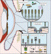
Functions of Membrane-Associated Mucins at the Human Ocular Surface. The graphic depicts some of the focus areas of this article. Left. Sagittal section of the eye and eyelids is represented, with epithelia highlighted in light pink. Goblet cells can be observed in the conjunctival epithelium (purple). Meibomian glands (yellow) can be found at the lids, with their ducts opening at the lid margin. Top right. Detail of the barrier functions of MAMs. Multiple MUC16 are showed protruding from the plasma membrane as the main elements of the glycocalyx barrier. LGALS3 (galectin-3) pentamers can be found interacting with mucin glycans. This barrier is responsible of the exclusion of different substances, such as the clinical dye rose bengal. Bacteria and viruses can be excluded also by shed mucins, as it is the case of MUC1, represented here. Mid right. Detail of the interactions of MAMs with elements of the cytoskeleton. MUC16 participates in the formation of membrane microplicae and tight junctions (represented here with the transmembrane protein occluding and ZO1 in blue). Some of these functions have been related to their interaction with actin filaments. Bottom right. Detail of the immunomodulatory functions of MAMs. A question mark is used to indicate functions that have not been demonstrated in the eye to date. Inhibitory effect on Toll-like receptors (fuchsia) has been described for MUC1 (left) and MUC16 (right) at the ocular surface, reducing the activation of NF-kappaB. Previous studies showed that MUC1 acted by inhibiting recruitment of MyD88. Studies in airways have demonstrated that MUC1-CT interacts with the glucocorticoid receptor (NR3C1), facilitating its migration to the nucleus, while MUC4 (right) can inhibit its translocation. Finally, studies in different systems have described interactions between MUC1-CT and different regulatory elements of the NF-kappaB pathway, although the final result of these interactions is not clear.
Figure 3.
Figure 3.
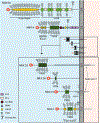
Modular Architecture of Membrane-Associated Mucins of the Human Ocular Surface. The extended conformation of each extracellular domain (ED) is to the left of the plasma membrane (gray bar) and each cytoplasmic tail (CT) to the right, both drawn to scale. The transmembrane domains are indicated as gray boxes embedded in the plasma membrane. MUC20 has been experimentally determined to associate with the plasma membrane, but has no transmembrane domain, and thus no CT has been identified (discussed in the text). Because of the extreme size differences between the large and small MAMs, an expanded view of the ED is shown for the small MAMs. Signal peptides are located at the amino-terminus of each protein (red blobs). The cleavage sites in the EDs are indicated by scissors.

Similar articles

Cited by

References

    1. Madison KC. Barrier function of the skin: “la raison d’etre” of the epidermis. The Journal of investigative dermatology. 2003;121(2):231–41. Epub 2003/07/26. doi: 10.1046/j.1523-1747.2003.12359.x. - DOI - PubMed
    1. Gipson IK. The ocular surface: the challenge to enable and protect vision: the Friedenwald lecture. Investigative ophthalmology & visual science. 2007;48(10):4390; 1–8. Epub 2007/09/28. doi: 10.1167/iovs.07-0770. - DOI - PMC - PubMed
    1. Hollingsworth MA, Swanson BJ. Mucins in cancer: protection and control of the cell surface. Nature reviews Cancer. 2004;4(1):45–60. doi: 10.1038/nrc1251. - DOI - PubMed
    1. Willcox MDP, Argueso P, Georgiev GA, Holopainen JM, Laurie GW, Millar TJ, et al. TFOS DEWS II Tear Film Report. The ocular surface. 2017;15(3):366–403. doi: 10.1016/j.jtos.2017.03.006. - DOI - PMC - PubMed
    1. Georgiev GA, Eftimov P, Yokoi N. Contribution of Mucins towards the Physical Properties of the Tear Film: A Modern Update. International journal of molecular sciences. 2019;20(24). Epub 2019/12/11. doi: 10.3390/ijms20246132. - DOI - PMC - PubMed

Publication types