Review
. 2021 May 22;46(1):37-49.
doi: 10.1247/csf.21015. Epub 2021 Mar 26.

Multiple Ways for Stress Sensing and Regulation of the Endoplasmic Reticulum-stress Sensors

Affiliations
Review

Multiple Ways for Stress Sensing and Regulation of the Endoplasmic Reticulum-stress Sensors

Quynh Giang Le et al. Cell Struct Funct. .

Abstract

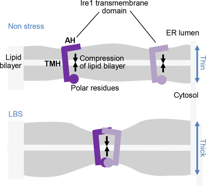
Dysfunction of the endoplasmic reticulum (ER), so-called ER stress, is accompanied with accumulation of unfolded proteins in the ER. Eukaryotic cells commonly have an ER-located transmembrane protein, Ire1, which triggers cellular protective events against ER stress. In animal cells, PERK and ATF6 also initiate the ER-stress response. As a common strategy to control the activity of these ER-stress sensors, an ER-resident molecular chaperone, BiP, serves as their negative regulator, and dissociates from them in response to ER stress. Although it sounds reasonable that unfolded proteins and Ire1 compete for BiP association, some publications argue against this competition model. Moreover, yeast Ire1 (and possibly also the mammalian major Ire1 paralogue IRE1α) directly detects ER-accumulated unfolded proteins, and subsequently oligomerizes for its further activation. Apart from protein misfolding, the saturation of membrane phospholipids is another outcome of ER-stressing stimuli, which is sensed by the transmembrane domain of Ire1. This review describes the canonical and up-to-date insights concerning stress-sensing and regulatory mechanisms of yeast Ire1 and metazoan ER-stress sensors.Key words: endoplasmic reticulum, stress, unfolded protein response, molecular chaperone.

Keywords: endoplasmic reticulum; molecular chaperone; stress; unfolded protein response.


Figures

Fig. 1
Fig. 1
Structure of Ire1 and PERK. Unlike those of the NUCR or the JIDR, the amino-acid sequences of the cLD are weakly but significantly conserved among these proteins. In our previous publication (Kimata et al., 2004), the NUCR, the cLD and the JIDR of the yeast Ire1 were named as Subregions I, II–IV and V.
Fig. 2
Fig. 2
Functions of yeast Ire1 and mammalian IRE1α. Self-association and auto-phosphorylation of Ire1 lead to its activation as RNase, which evokes the indicated events. While, as described later in this article, Ire1 is more strongly activated as high-order oligomers, here it is presented as dimers. Although not shown in this figure, it is also likely that IRE1α phosphorylates other proteins.
Fig. 3
Fig. 3
Our model for upregulation of Ire1 upon ER accumulation of unfolded proteins in yeast cells. Under non-stress conditions, the JIDR (the BiP-binding site) and the NUCR inhibit the self-association of Ire1. Upon ER stress, BiP is dissociated from Ire1, which is then dimerized. The Ire1 dimers are automatically phosphorylated and then captures ADP for their activation as RNase. Meanwhile, the Ire1 dimers directly captures ER-accumulated unfolded proteins and are further bundled, leading to more potent UPR.
Fig. 4
Fig. 4
Possible involvement of the Ire1 transmembrane-domain structure in the activation of Ire1 upon LBS. The short TMH of Ire1 is juxtaposed N-terminally to the AH and C-terminally to polar residues, causing compression of the lipid bilayer (Halbleib et al., 2017; Covino et al., 2018). When the lipid-bilayer structure is disturbed, for example, through membrane-lipid saturation, free energy cost for the lipid-bilayer compression by the Ire1 transmembrane domain is increased. Ire1 molecules are then gathered to cope with this situation.
Fig. 5
Fig. 5
Relationship between BiP and Ire1. (A) In general, the HSP70-family molecular chaperones capture client substrate proteins dependently on its nucleotide-binding states. (B) and (C) As described in the main text, two theories are proposed to explain the association/dissociation mode between IRE1α and ER-accumulated unfolded proteins. Nucleotides bound to BiP are not shown in panels B and C.
Fig. 6
Fig. 6
A possible model to explain how IRE1α is activated upon ER accumulation of unfolded proteins in animal cells. Under non-stress conditions, BiP is bound to the cLD and JIDR of IRE1α, which then stays non-self-associated. Upon ER accumulation of unfolded proteins, IRE1α dissociates from BiP and forms homo-dimers. It is uncertain if IRE1α and unfolded proteins bind to the same site of BiP. Similarly to those of yeast Ire1, IRE1αdimers are further bundled through their direct interaction with unfolded proteins. As described in the main text, it is possible that IRE1α dimers and oligomers have distinct biological functions.
Fig. 7
Fig. 7
Activation process of ATF6. In non-stressed cells, ATF6 is retained in the ER as BiP-bound and oxidized forms. Upon ER stress, ATF6 dissociates from BiP and is reduced (cleavage of the disulfide bonds). The monomeric ATF6 proteins are then transported to the Golgi apparatus and proteolytically cleaved.

Similar articles

Cited by

References

    1. Ali, M.M.U., Bagratuni, T., Davenport, E.L., Nowak, P.R., Silva-Santisteban, M.C., Hardcastle, A., McAndrews, C., Rowlands, M.G., Morgan, G.J., Aherne, W., Collins, I., Davies, F.E., and Pearl, L.H.. 2011. Structure of the Ire1 autophosphorylation complex and implications for the unfolded protein response. EMBO J., 30: 894–905. - PMC - PubMed
    1. Amin-Wetzel, N., Saunders, R.A., Kamphuis, M.J., Rato, C., Preissler, S., Harding, H.P., and Ron, D.. 2017. A J-Protein Co-chaperone Recruits BiP to Monomerize IRE1 and Repress the Unfolded Protein Response. Cell, 171: 1625–1637. - PMC - PubMed
    1. Amin-Wetzel, N., Neidhardt, L., Yan, Y., Mayer, M.P., and Ron, D.. 2019. Unstructured regions in IRE1alpha specify BiP-mediated destabilisation of the luminal domain dimer and repression of the UPR. eLife, 8: e.50793. - PMC - PubMed
    1. Aragón, T., van Anken, E., Pincus, D., Serafimova, I.M., Korennykh, A.V., Rubio, C.A., and Walter, P.. 2009. Messenger RNA targeting to endoplasmic reticulum stress signalling sites. Nature, 457: 736–740. - PMC - PubMed
    1. Bashir, S., Banday, M., Qadri, O., Bashir, A., Hilal, N., Nida-I-Fatima, Rader, S., and Fazili, K.M.. 2020. The molecular mechanism and functional diversity of UPR signaling sensor IRE1. Life Sci., 265: 118740. - PubMed

MeSH terms

Substances