

Progression of CIN1/LSIL HPV Persistent of the Cervix: Actual Progression or CIN3 Coexistence
- PMID: 33776406
- PMCID: PMC7972837
- DOI: 10.1155/2021/6627531
Progression of CIN1/LSIL HPV Persistent of the Cervix: Actual Progression or CIN3 Coexistence
Abstract
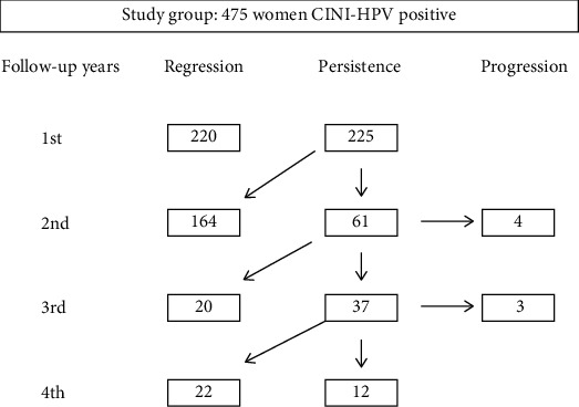
Objective: The natural history of the CIN1 lesions is characterized by an elevated rate of spontaneous regression (80%), some authors recognize a capacity to progress to HSIL in 10% of cases, and other authors do not recognize the capacity of progression of LSIL (CIN1). This study was aimed to evaluate the incidence of progression to HSIL (CIN3) in women with a histological diagnosis of LSIL (CIN1). Furthermore, to this end, we studied the histological outcomes of cone specimens collected by the LEEP.
Methods: All the data were retrospectively analyzed. All participants underwent a follow-up of 4 years, during which each woman underwent an HPV test and genotyping, cervical cytological sampling, or biopsy every six months. The endpoint was the histological confirmation of CIN3 lesions in any moment during follow-up.
Results: Progression to CIN3 occurred in 7 cases (1,5%). Analyzing the histological exams of the cones of the 7 cases that progressed to CIN3, we found the coexistence of CIN1 and CIN3 lesions in all cases.
Conclusion: After 4 years of follow-up, only 1.5% (7/475) of the women with LSIL developed CIN3, all within the first two years of follow-up, and were immediately treated. The most likely explanations for "progression" from LSIL to HSIL are (1) actual progression, (2) underdiagnosis of HSIL on initial biopsy, (3) overdiagnosis of HSIL on follow-up biopsy/cone, and (4) CIN3 arose de novo. Analyzing the histological exams of the cones of the 7 cases that progressed to high-grade, we found the coexistence of CIN1 and CIN3 lesions in all cases. Some recent studies have shown that a viral genotype corresponds to different lesions in the same cervix; therefore, CIN1 coexisting with CIN3 does not always indicate progression of CIN1. Other authors have doubted the capacity of LSIL to progress.
Copyright © 2021 Maria Teresa Bruno et al.
Conflict of interest statement
The authors declare that they have no conflict of interests.
Figures
References
-
- Darragh T. M., Colgan T. J., Cox J. T., et al. The Lower Anogenital Squamous Terminology Standardization Project for HPV-Associated Lesions: background and consensus recommendations from the College of American Pathologists and the American Society for Colposcopy and Cervical Pathology. Journal of Lower Genital Tract Disease. 2012;16(3):205–242. doi: 10.1097/LGT.0b013e31825c31dd. - DOI - PubMed
-
- Italian society of colposcopy and cervicovaginal pathology. Guidelines for the management of abnormal pap smear. Società Italiana di Colposcopia e Patologia Cervico Vaginale. 2006;1:1–2.
MeSH terms
LinkOut - more resources
Full Text Sources
Other Literature Sources
Medical
