Review
. 2021 Mar 18:13:303-328.
doi: 10.2147/JEP.S267383. eCollection 2021.

Advances in Our Understanding of the Molecular Mechanisms of Action of Cisplatin in Cancer Therapy

Affiliations
Review

Advances in Our Understanding of the Molecular Mechanisms of Action of Cisplatin in Cancer Therapy

Paul B Tchounwou et al. J Exp Pharmacol. .

Abstract

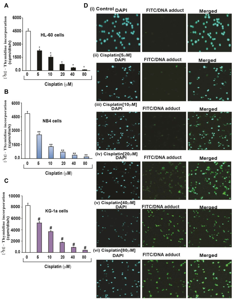
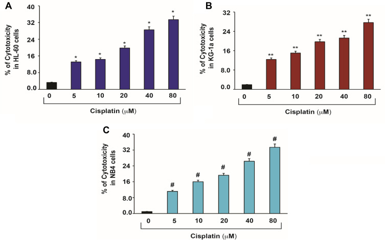
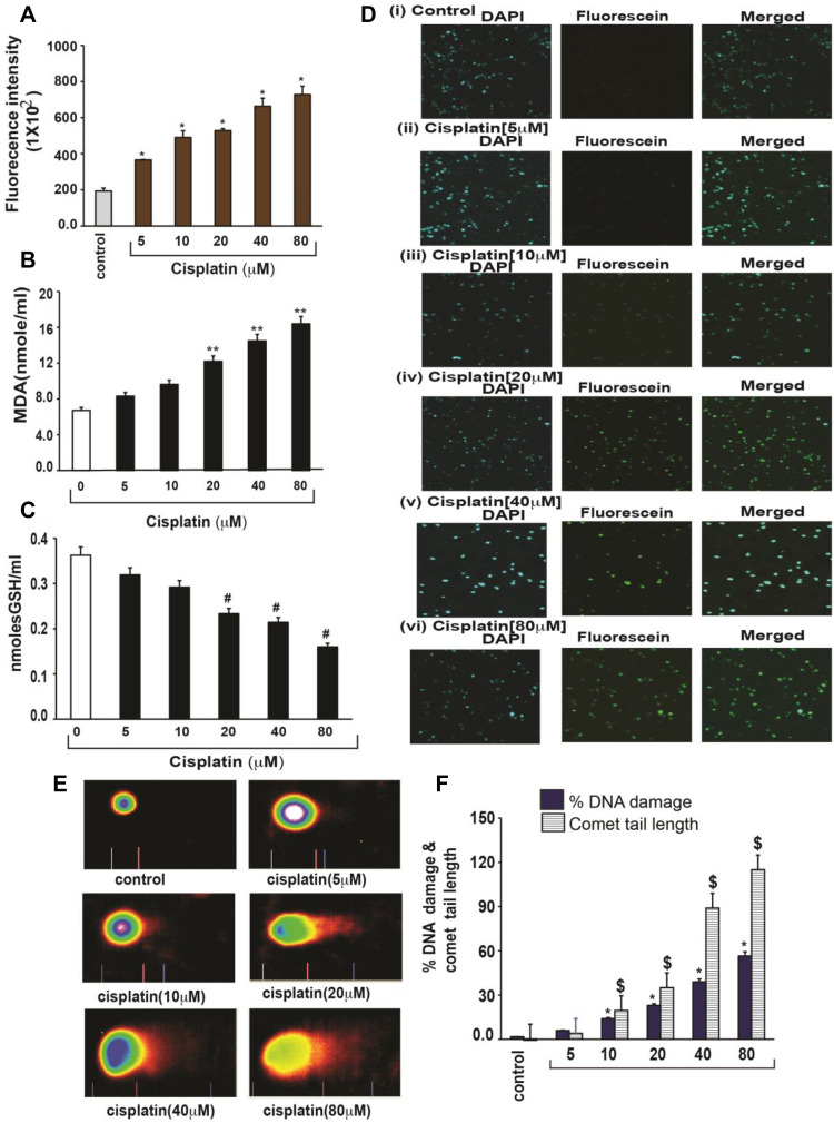
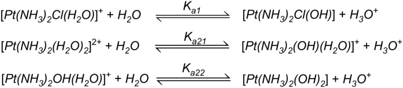
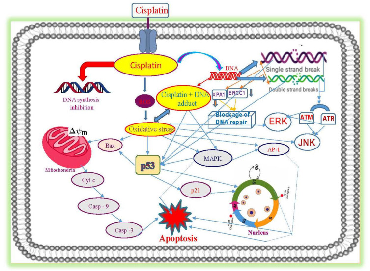
Cisplatin and other platinum-based chemotherapeutic drugs have been used extensively for the treatment of human cancers such as bladder, blood, breast, cervical, esophageal, head and neck, lung, ovarian, testicular cancers, and sarcoma. Cisplatin is commonly administered intravenously as a first-line chemotherapy for patients suffering from various malignancies. Upon absorption into the cancer cell, cisplatin interacts with cellular macromolecules and exerts its cytotoxic effects through a series of biochemical mechanisms by binding to Deoxyribonucleic acid (DNA) and forming intra-strand DNA adducts leading to the inhibition of DNA synthesis and cell growth. Its primary molecular mechanism of action has been associated with the induction of both intrinsic and extrinsic pathways of apoptosis resulting from the production of reactive oxygen species through lipid peroxidation, activation of various signal transduction pathways, induction of p53 signaling and cell cycle arrest, upregulation of pro-apoptotic genes/proteins, and down-regulation of proto-oncogenes and anti-apoptotic genes/proteins. Despite great clinical outcomes, many studies have reported substantial side effects associated with cisplatin monotherapy, while others have shown substantial drug resistance in some cancer patients. Hence, new formulations and several combinational therapies with other drugs have been tested for the purpose of improving the clinical utility of cisplatin. Therefore, this review provides a comprehensive understanding of its molecular mechanisms of action in cancer therapy and discusses the therapeutic approaches to overcome cisplatin resistance and side effects.

Keywords: cancer treatment; cisplatin; combination therapy; molecular mechanisms of action.


Conflict of interest statement

All authors have declared that they do not have any competing and/or financial interests for this work.

Figures

Figure 1
Figure 1
Cisplatin-induced growth inhibition and formation of DNA-adduct in APL cells. APL cells (HL-60, NB4 and KG-1a) were exposed to various concentrations (0, 5, 10, 20, 40, and 80 μM) of cisplatin for 24 hours and further incubated for 24 hours with tritium labeled thymidine. After incubation, cells were harvested by centrifugation and counted using liquid scintillation analyzer. 3H-methyl thymidine incorporation was expressed as cpm/dish. Data represent the means of three independent experiments ± SDs. Highly statistically significant decreases (p < 0.01) in cell proliferation were observed in all cisplatin-treated APL cells including HL-60 (A), NB4 (B) and Kg-1a (C) cells. Cisplatin–induced formation of DNA adduct was assessed by immunocytochemistry and confocal microscopy analysis. APL cells were treated with various concentrations of cisplatin for 48 hours and immunocytochemistry as well as confocal microscopy using FITC filter were performed to confirm DNA adduct formation. The results showed that cisplatin caused a significant concentration-dependent increase in DNA-adduct formation in APL cells [D (i–vi)]. Multiple symbol indicators (*, **, #) indicate highly significant reductions (p<0.01) in growth between control and cisplatin-treated cells, respectively, for HL-60, NB4, and KG1a cells. Reproduced from Kumar S, Tchounwou PB. Molecular mechanisms of cisplatin cytotoxicity in acute promyelocytic leukemia cells. Oncotarget. 2015;6(38):40734–40746. Creative Commons license and disclaimer available from: http://creativecommons.org/licenses/by/4.0/legalcode.14
Figure 2
Figure 2
Cisplatin-induced cytotoxic effects in APL cells. APL cells were exposed to various concentrations (0, 5, 10, 20, 40 and 80 μM) of cisplatin for 48 hours and LDH released in medium was measured using Promega non-radioactive cytotoxicity assay technical bulletin protocol. Then, % cytotoxicity was calculated by dividing the levels of released LDH in treated cells over the total LDH released from control cells. Highly statistically significant increases (p < 0.01) in cytotoxicity were observed in all cisplatin-treated APL cells including HL-60 (A), KG-1a (B) and NB4 (C) cells in a concentration-dependent fashion. Multiple symbol indicators (*, **, #) indicate highly significant increases (p<0.01) in cytotoxicity between control and cisplatin-treated cells, respectively, for HL-60, NB4, and KG1a cells.  Reproduced from Kumar S, Tchounwou PB. Molecular mechanisms of cisplatin cytotoxicity in acute promyelocytic leukemia cells. Oncotarget. 2015;6(38):40734–40746. Creative Commons license and disclaimer available from: http://creativecommons.org/licenses/by/4.0/legalcode.14
Figure 3
Figure 3
Cisplatin-induced oxidative stress and DNA damage in APL cells. APL cells were exposed to various concentrations (0, 5, 10, 20, 40, and 80 μM) of cisplatin for 48 hours. ROS release was assessed by spectrofluorometry based on DCF fluorescence intensity after 30 min incubation with dichlorofluorescein diacetate (DCFDA). After incubation, ROS release was measured through measuring DCF fluorescence intensity by spectrofluorometry. Malondialdehyde (MDA) and GSH concentration levels were also measured by spectrophotometry. DNA damage was analyzed by both TUNEL and alkaline gel electrophoresis (Comet) assays. (A) ROS concentrations; (B) MDA concentrations; (C) GSH concentrations; (D) TUNEL assay images of DNA damage; (E) Comet assay images of DNA damage; (F) Percentages of DNA damage, and Comet tail lengths. Multiple symbol indicators (*, **, #, $) indicate highly significant increases (p<0.01) in ROS (*), MDA (**), DNA damage (*) and Comet tail length ($), and highly significant decreases (p<0.01) in GSH ($) between control and cisplatin-treated cells.  Reproduced from Kumar S, Tchounwou PB. Molecular mechanisms of cisplatin cytotoxicity in acute promyelocytic leukemia cells. Oncotarget. 2015;6(38):40734–40746. Creative Commons license and disclaimer available from: http://creativecommons.org/licenses/by/4.0/legalcode.14
Figure 4
Figure 4
Chemical reactions showing hydrolysis of cisplatin. Reproduced with permission from  Burda JV, Zeizinger M, Leszczynski J. Hydration process as an activation of trans- and cisplatin complexes in anticancer treatment. DFT and Ab initio computational study of thermodynamic and kinetic parameters. J Comput Chem. 2005;26(9):907–914. Copyright © 2005 Wiley Periodicals, Inc.
Figure 5
Figure 5
Molecular mechanisms of cisplatin-induced cytotoxicity in APL cells.  Reproduced from Kumar S, Tchounwou PB. Molecular mechanisms of cisplatin cytotoxicity in acute promyelocytic leukemia cells. Oncotarget. 2015;6(38):40734–40746. Creative Commons license and disclaimer available from: http://creativecommons.org/licenses/by/4.0/legalcode.

References

    1. Alderden RA, Hall MD, Hambley TW. The discovery and development of cisplatin. J Chem Educ. 2006;83(5):728. doi:10.1021/ed083p728 - DOI
    1. Muggia FM, Bonetti A, Hoeschele JD, Rozencweig M, Howell SB. Platinum antitumor complexes: 50 Years since Barnett Rosenberg’s discovery. J Clin Oncol. 2015;33(35):4219–4226. doi:10.1200/JCO.2015.60.7481 - DOI - PubMed
    1. Rosenberg B, Van Camp L, Krigas T. Inhibition of cell division in Escherichia coli by electrolysis products from a platinum electrode [17]. Nature. 1965;205(4972):698–699. doi:10.1038/205698a0 - DOI - PubMed
    1. IARC, International Agency for Research on Cancer. IARC monographs on the evaluation of carcinogenic risks to humans overall evaluations of carcinogenicity: an updating of IARC monographs volumes 1 to 42; SUPPLEMENT 7. IARC Monogr Eval Carcinog RISKS TO HUMANS - Overall Eval Carcinog An Update IARC Monographs; 1987. - PubMed
    1. The Department of Chemistry at the University of Akron. Akron the chemical database; 2009. Available from: https://pubchem.ncbi.nlm.gov/compound/cis-Platin . Accessed March 04, 2021.