Case Reports
. 2021 Mar;12(1):41-45.
doi: 10.1159/000512304. Epub 2020 Dec 15.

Prenatal Diagnosis of Acromelic Frontonasal Dysostosis

Affiliations
Case Reports

Prenatal Diagnosis of Acromelic Frontonasal Dysostosis

Cristina Martínez-Payo et al. Mol Syndromol. 2021 Mar.

Abstract

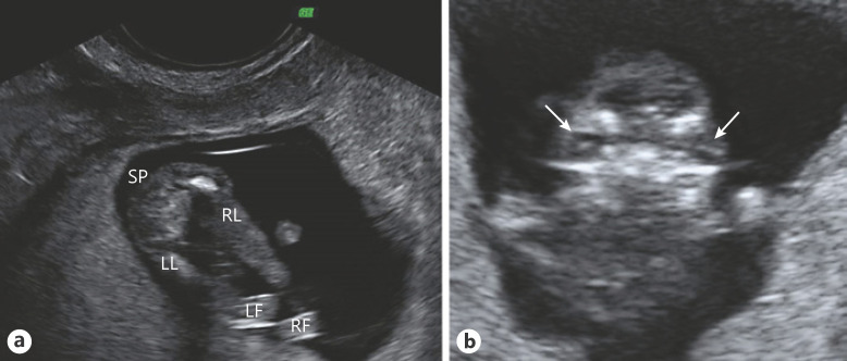
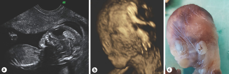
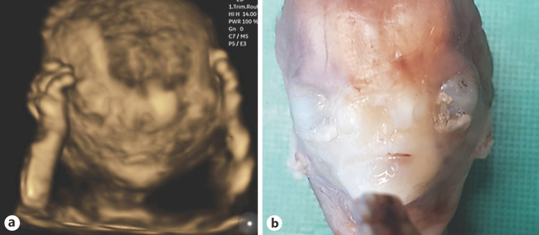
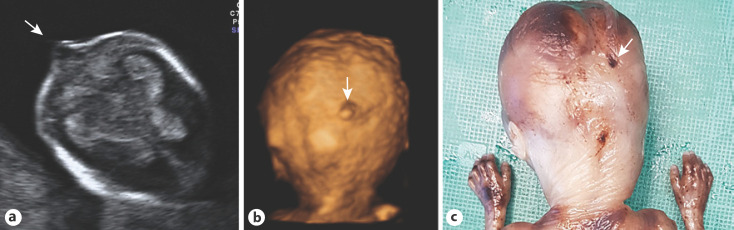
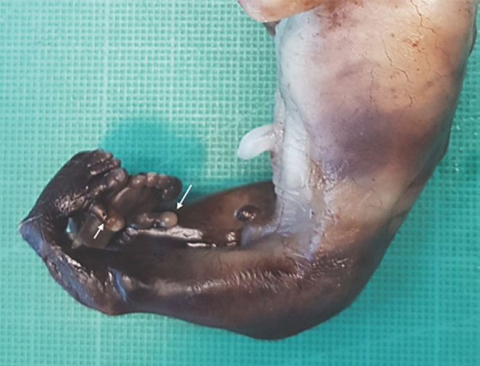
Acromelic frontonasal dysostosis (AFND; MIM #603671) is a rare autosomal dominant genetic disorder caused by a heterozygous mutation in the ZSWIM6 (KIAA1577) gene located at chromosome 5q12.1. It is phenotypically characterized by frontonasal malformation with hypertelorism, telecanthus, nasal clefting or bifid nasal tip, wide fontanels and sutures, brachycephaly, and cleft palate. The patients also present with central nervous system malformations such as encephalocele, agenesis of the corpus callosum, or interhemispheric lipoma. Limb malformations can also be found, including preaxial polydactyly of the feet and sometimes postaxial polydactyly of the hands, talipes equinovarus, or tibia malformations. Here, we present a case of early prenatal diagnosis of AFND with ultrasound and necropsy images which show the phenotypic findings of this syndrome.

Keywords: Acromelic frontonasal dysostosis; Central nervous system malformation; Craniofacial malformation; Limb malformation; ZSWIM6.


Conflict of interest statement

The authors have no conflicts of interest to declare.

Figures

Fig. 1
Fig. 1
Ultrasound images at the 13th week of pregnancy. a Both lower limbs of the fetus. Note mesomelic shortening and malposition of both feet. b Coronal view of the fetal facies. The arrows point to both crystalline lens, showing pronounced hypertelorism. SP, spine; LL, left leg; RL, right leg; LF, left foot; RF, right foot.
Fig. 2
Fig. 2
a Abnormal ultrasound image of the fetal profile at the 15th week of pregnancy with a flat profile and an absent nose. b 3D ultrasound reconstruction of the fetal head. The flat profile and the absent nose can be seen. Note nasal clefting. c Necropsy image additionally shows low-set ears.
Fig. 3
Fig. 3
a 3D reconstruction of the fetal facies at the 15th week of pregnancy showing upper limbs on the side the head. Pronounced hypertelorism, as well as an abnormal development of the frontonasal area, with an undefined bifid nose are shown. b Fetal necropsy image demonstrating the same findings.
Fig. 4
Fig. 4
a Ultrasound image of the fetal head at the 15th week of pregnancy, in a transversal cut in transthalamic plane, which shows a small encephalocele (arrow). The 3D ultrasound reconstruction (b) and the photographic image of the fetal necropsy (c) show the fetal occipital area, and the arrows point to the small encephalocele. Note normal fetal hands.
Fig. 5
Fig. 5
Fetal necropsy showing the malformation of the lower limbs affecting the mesomelic segment and both feet as well as bilateral preaxial polydactyly.

References

    1. Palmer EE, Kumar R, Gordon CT, Shaw M, Hubert L, Carroll R, et al. A Recurrent De Novo Nonsense Variant in ZSWIM6 Results in Severe Intellectual Disability without Frontonasal or Limb Malformations. Am J Hum Genet. 2017;101((6)):995–1005. - PMC - PubMed
    1. Richards S, Aziz N, Bale S, Bick D, Das S, Gastier-Foster J, et al. Standards and guidelines for the interpretation of sequence variants: a joint consensus recommendation of the American College of Medical Genetics and Genomics and the Association for Molecular Pathology. Genet Med. 2015;17((5)):405–24. - PMC - PubMed
    1. Sedano HO, Cohen MM, Jr, Jirasek J, Gorlin RJ. Frontonasal dysplasia. J Pediatr. 1970;76((6)):906–13. - PubMed
    1. Smith JD, Hing AV, Clarke CM, Johnson NM, Perez F, Park SS, et al. Exome sequencing identifies a recurrent de novo ZSWIM6 mutation associated with acromelic frontonasal dysostosis. Am J Hum Genet. 2014;95:235–40. - PMC - PubMed
    1. Twigg SR, Ousager LB, Miller KA, Zhou Y, Elalaoui SC, Sefiani A, et al. Acromelic frontonasal dysostosis and ZSWIM6 mutation: phenotypic spectrum and mosaicism. Clin Genet. 2016;90((3)):270–5. - PMC - PubMed

Publication types