Review
. 2021 Mar 10:15:614412.
doi: 10.3389/fnins.2021.614412. eCollection 2021.

Circuit Mechanisms of L-DOPA-Induced Dyskinesia (LID)

Affiliations
Review

Circuit Mechanisms of L-DOPA-Induced Dyskinesia (LID)

Kai Yang et al. Front Neurosci. .

Abstract

L-DOPA is the criterion standard of treatment for Parkinson disease. Although it alleviates some of the Parkinsonian symptoms, long-term treatment induces L-DOPA-induced dyskinesia (LID). Several theoretical models including the firing rate model, the firing pattern model, and the ensemble model are proposed to explain the mechanisms of LID. The "firing rate model" proposes that decreasing the mean firing rates of the output nuclei of basal ganglia (BG) including the globus pallidus internal segment and substantia nigra reticulata, along the BG pathways, induces dyskinesia. The "firing pattern model" claimed that abnormal firing pattern of a single unit activity and local field potentials may disturb the information processing in the BG, resulting in dyskinesia. The "ensemble model" described that dyskinesia symptoms might represent a distributed impairment involving many brain regions, but the number of activated neurons in the striatum correlated most strongly with dyskinesia severity. Extensive evidence for circuit mechanisms in driving LID symptoms has also been presented. LID is a multisystem disease that affects wide areas of the brain. Brain regions including the striatum, the pallidal-subthalamic network, the motor cortex, the thalamus, and the cerebellum are all involved in the pathophysiology of LID. In addition, although both amantadine and deep brain stimulation help reduce LID, these approaches have complications that limit their wide use, and a novel antidyskinetic drug is strongly needed; these require us to understand the circuit mechanism of LID more deeply.

Keywords: L-DOPA induced dyskinesia; Parkinson’s disease; firing pattern; firing rate; neuronal oscillation.


Conflict of interest statement

The authors declare that the research was conducted in the absence of any commercial or financial relationships that could be construed as a potential conflict of interest.

Figures

FIGURE 1
FIGURE 1
The direct and indirect pathway of the BG. In the health, the activation of the direct pathway reduces the output of the BG, stimulating the thalamus and cortex, which promotes the movement. While the stimulation of the indirect pathway increases the BG output, suppressing the cortex and inhibiting the movement. In PD, the deficiency of DA in the striatum causes the overactivation of indirect pathway, as well as the hypoactivation of direct pathway, resulting in the suppression of the thalamus and the cortex, which inhibits the movement. In contrast, in LID, the activity of direct output pathway is increased while the indirect output pathway is inhibited, which disinhibits the thalamus and the cortex, resulting in the increase of the movement. Red lines indicate excitatory transmission, whereas blue lines mean inhibitory transmission, and line width represents the strength of neuronal transmission. This figure adapted from McGregor and Nelson (2019).
FIGURE 2
FIGURE 2
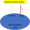
Oscillation model of LID. Oscillation model proposes that dyskinesia is characterized by significant changes in oscillations; usually β oscillation is reduced (Delaville et al., 2015), whereas γ oscillation is enhanced (Halje et al., 2012).
FIGURE 3
FIGURE 3
Brain regions involved in the pathophysiology of LID. Besides the striatum, other brain regions including the pallidal–subthalamic network, the motor cortex, the thalamus, the cerebellum, the dorsal raphe nucleus (DRN), the LC, the LHb, and the BNST are also involved in LID. During LID, LHb neurons are suggested to induce the aberrant DA release in the striatum from serotonergic terminals via DRN and generate LID symptoms (Carta et al., 2007; Bernard and Veh, 2012), but the mechanisms how the LC and the BNST affect LID remain unknown (red “?” represents unknown). In addition, the BG and the cerebellum are thought to communicate at the subcortical level. The subthalamic nucleus (STN) sends projections to the cerebellar cortex through pontine nuclei (PN) (blue line) (Bostan et al., 2010), whereas the dentate nucleus in the cerebellum sends connection to the striatum via the thalamus (yellow line) (Hoshi et al., 2005).

References

    1. Alachkar A., Brotchie J. M., Jones O. T. (2012). Changes in the mRNA levels of alpha2A and alpha2C adrenergic receptors in rat models of Parkinson’s disease and L-DOPA-induced dyskinesia. J. Mol. Neurosci. 46 145–152. 10.1007/s12031-011-9539-x - DOI - PubMed
    1. Alam M., Capelle H. H., Schwabe K., Krauss J. K. (2014). Effect of deep brain stimulation on levodopa-induced dyskinesias and striatal oscillatory local field potentials in a rat model of Parkinson’s disease. Brain Stimul. 7 13–20. 10.1016/j.brs.2013.09.001 - DOI - PubMed
    1. Alberico S. L., Kim Y. C., Lence T., Narayanan N. S. (2017). Axial levodopa-induced dyskinesias and neuronal activity in the dorsal striatum. Neuroscience 343 240–249. 10.1016/j.neuroscience.2016.11.046 - DOI - PMC - PubMed
    1. Alegre M., Alonso-Frech F., Rodriguez-Oroz M. C., Guridi J., Zamarbide I., Valencia M., et al. (2005). Movement-related changes in oscillatory activity in the human subthalamic nucleus: ipsilateral vs. contralateral movements. Eur. J. Neurosci. 22 2315–2324. 10.1111/j.1460-9568.2005.04409.x - DOI - PubMed
    1. Allers K. A., Bergstrom D. A., Ghazi L. J., Kreiss D. S., Walters J. R. (2005). MK801 and amantadine exert different effects on subthalamic neuronal activity in a rodent model of Parkinson’s disease. Exp. Neurol. 191 104–118. 10.1016/j.expneurol.2004.08.030 - DOI - PubMed

LinkOut - more resources