. 2021 Mar 10:11:612766.
doi: 10.3389/fimmu.2020.612766. eCollection 2020.

Reduction of Allergic Lung Disease by Mucosal Application of Toxoplasma gondii-Derived Molecules: Possible Role of Carbohydrates

Affiliations

Reduction of Allergic Lung Disease by Mucosal Application of Toxoplasma gondii-Derived Molecules: Possible Role of Carbohydrates

Elke Korb et al. Front Immunol. .

Abstract

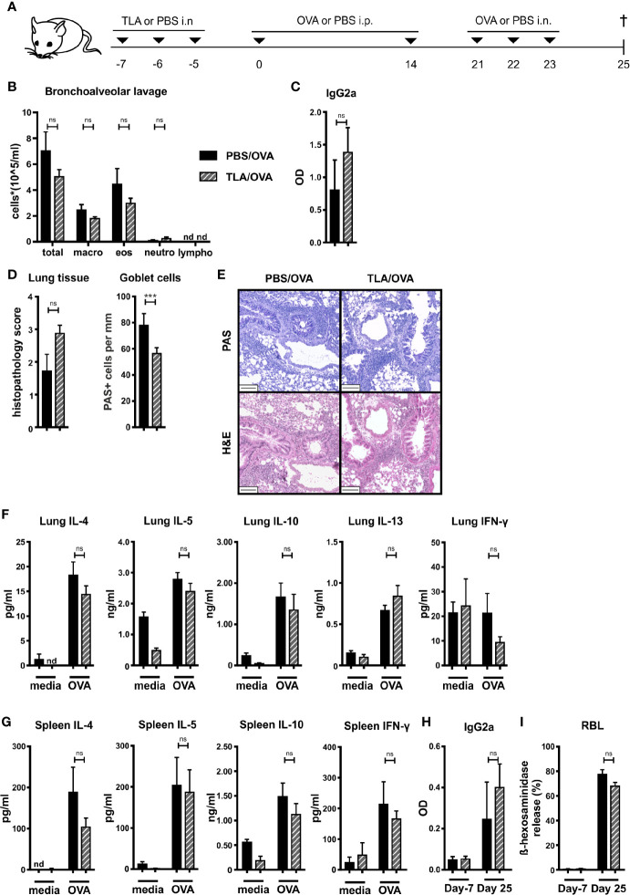
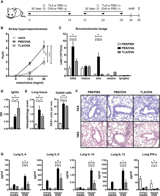
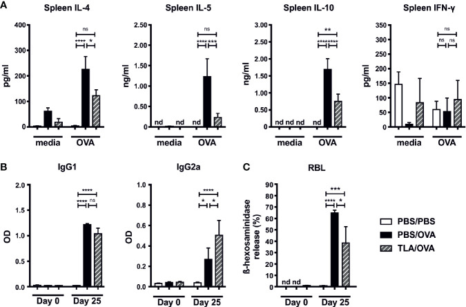
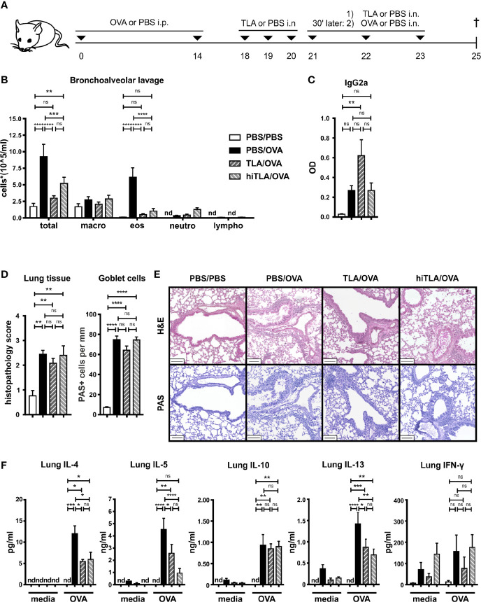
Background: The hygiene hypothesis suggests a link between parasitic infections and immune disorders, such as allergic diseases. We previously showed that infection with Toxoplasma gondii or systemic application of T. gondii tachyzoites lysate antigen (TLA) in a prophylactic, but not therapeutic protocol, prevented allergic airway inflammation in mice. Here we tested the effect of prophylactic and therapeutic application of TLA via the mucosal route.

Methods: Mice were intranasally treated with TLA either i) prior to sensitization, ii) during sensitization and challenge, or iii) after sensitization with ovalbumin (OVA). Recruitment of inflammatory cells to the lung, cytokine levels in restimulated lung and spleen cell cultures as well as levels of OVA-specific antibodies in serum were measured. In parallel, the effect of native TLA, heat-inactivated (hiTLA) or deglycosylated TLA (dgTLA) on sensitized splenocytes was evaluated ex vivo.

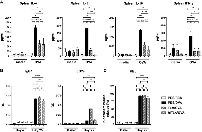
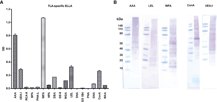
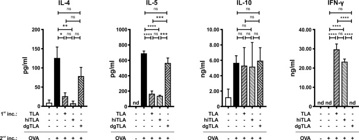
Results: When applied together with OVA i) during systemic sensitization and local challenge or ii) exclusively during local challenge, TLA reduced infiltration of eosinophils into the lung, OVA-specific type 2 cytokines in restimulated lung cell cultures, and partially, type 2 cytokines in restimulated spleen cell cultures in comparison to allergic controls. No beneficial effect was observed when TLA was applied prior to the start of sensitization. Analysis of epitope sugars on TLA indicated a high abundance of mannose, fucose, N-acetylglucosamine, and N-acetylgalactosamine. Deglycosylation of TLA, but not heat-inactivation, abolished the potential of TLA to reduce type 2 responses ex vivo, suggesting a significant role of carbohydrates in immunomodulation.

Conclusion: We showed that mucosal application of TLA reduced the development of experimental allergy in mice. The beneficial effects depended on the timing of the application in relation to the time point of sensitization. Not only co-application, but also therapy in sensitized/allergic animals with native TLA reduced local allergic responses. Furthermore, we show that TLA is highly glycosylated and glycoconjugates seem to play a role in anti-allergic effects. In summary, given the powerful modulatory effect that TLA exhibits, understanding its exact mechanisms of action may lead to the development of novel immunomodulators in clinical application.

Keywords: Toxoplasma gondii; allergic airway inflammation; carbohydrates; deglycosylation; hygiene hypothesis; immunomodulation; parasites; tachyzoites lysate antigen.


Conflict of interest statement

The authors declare that the research was conducted in the absence of any commercial or financial relationships that could be construed as a potential conflict of interest.

Figures

Figure 1
Figure 1
Prophylactic treatment with TLA fails to prevent allergic airway inflammation. (A) Experimental design. (B) Differential cell counts in bronchoalveolar lavage (BALF). (C) Levels of OVA-specific antibody IgG2a in BALF collected at the end of the experiment. (D) Average histopathology score and number of Periodic acid-Schiff (PAS)-positive goblet cells of lung sections. (E) PAS or hematoxylin and eosin (H&E)-stained lung sections from 1 representative example from each group (n = 5); scale bars, 100 µm. (F) Levels of IL-4, IL-5, IL-10, IL-13, and IFN-γ after medium and ovalbumin (OVA) restimulation of lung cells. (G) Levels of IL-4, IL-5, IL-10, and IFN-γ after medium and OVA restimulation of spleen cells. (H) Levels of OVA-specific antibody IgG2a in serum collected at the beginning and at the end of the experiment. (I) Release of β-hexosaminidase by rat basophil leukemia (RBL) cells. Graphs show results from 1 representative experiment from 2 independent experiments with 5 mice per group ( Figures 1B–I ). Error bars show mean ± SEM. TLA, tachyzoites lysate antigen; OVA, ovalbumin; i.n., intranasal; i.p., intraperitoneal; macro, macrophages; eos, eosinophils; neutro, neutrophils; lympho, lymphocytes; OD, optical density; nd, not detectable; ns, not significant; ***P < 0.001.
Figure 2
Figure 2
TLA reduces allergic airway inflammation in a co-application model. (A), Experimental design. (B) Airway hyperresponsiveness in response to methacholine. (C) Differential cell count in bronchoalveolar lavage (BALF). (D) Levels of OVA-specific antibody IgG2a in BALF collected at the end of the experiment. (E) Average histopathology score and number of Periodic acid-Schiff (PAS)-positive goblet cells of lung sections. (F) PAS or hematoxylin and eosin (H&E)-stained lung sections from 1 representative example from each group (n = 5); scale bars, 100 µm. (G) Levels of IL-4, IL-5, IL-10, IL-13, and IFN-γ after medium and OVA restimulation of lung cells. Graphs show results from 1 experiment with 5 mice per group ( Figure 2B , PBS/PBS group in Figures 2C–G ) or from 1 representative experiment from 2 independent experiments with 5 mice per group (groups PBS/OVA and TLA/OVA, Figures 2C–G ). Error bars show mean ± SEM. TLA, tachyzoites lysate antigen; OVA, ovalbumin; i.n., intranasal; i.p., intraperitoneal; macro, macrophages; eos, eosinophils; neutro, neutrophils; lympho, lymphocytes; nd, not detectable; ns, not significant; OD, optical density; *P < 0.05; **P < 0.01; ***P < 0.001; ****P < 0.0001.
Figure 3
Figure 3
TLA reduces systemic IL-4 and serum IgE-levels in a co-application model. (A) Levels of IL-4, IL-5, IL-10, and IFN-γ after medium and ovalbumin (OVA) restimulation of spleen cells from mice treated as in Figure 2A . (B) Levels of OVA-specific antibodies IgG1 and IgG2a in serum collected at the beginning and at the end of the experiment. (C) Release of β-hexosaminidase by rat basophil leukemia (RBL) cells. Graphs show results from 1 experiment with 5 mice per group (PBS/PBS) or 1 representative experiment from 2 independent experiments with 5 mice per group (PBS/OVA and TLA/OVA). Error bars show mean ± SEM. TLA, tachyzoites lysate antigen; OD, optical density; nd, not detectable; ns, not significant; *P < 0.05; **P < 0.01; ***P < 0.001; ****P < 0.0001.
Figure 4
Figure 4
Therapeutic treatment with native or heat-inactivated TLA reduces allergic airway inflammation. (A) Experimental design. (B) Differential cell count in bronchoalveolar lavage (BALF). (C) Levels of OVA-specific antibody IgG2a in BALF collected at the end of the experiment. (D) Average histopathology score and number of Periodic acid-Schiff (PAS)-positive goblet cells of lung sections. (E) PAS or H&E-stained lung sections from 1 representative example from each group (n = 5); scale bars, 100 µm. (F) Levels of IL-4, IL-5, IL-10, IL-13, and IFN-γ after medium and OVA restimulation of lung cells. Graphs show results from 1 representative experiment from 2 independent experiments with 5 mice per group. Error bars show mean ± SEM. TLA, tachyzoites lysate antigen; hiTLA, heat-inactivated; OVA, ovalbumin; i.n., intranasal; i.p., intraperitoneal; macro, macrophages; eos, eosinophils; neutro, neutrophils; lympho, lymphocytes; n.s., not significant; OD, optical density; *P < 0.05; **P < 0.01; ***P < 0.001; ****P < 0.0001.
Figure 5
Figure 5
Therapeutic treatment with native or heat-inactivated TLA reduces systemic type 2 responses. (A) Levels of IL-4, IL-5, IL-10, and IFN-γ after medium and ovalbumin (OVA) restimulation of spleen cells from mice treated as in Figure 4A . (B) Levels of OVA-specific antibodies IgG1 and IgG2a in serum collected at the beginning and at the end of the experiment. (C) Release of β-hexosaminidase by rat basophil leukemia (RBL) cells. Graphs show results from 1 representative experiment from 2 independent experiments with 5 mice per group. Error bars show mean ± SEM. nd, not detectable; TLA, tachyzoites lysate antigen; hiTLA, heat-inactivated TLA; ns, not significant; OD, optical density; *P < 0.05; **P < 0.01; ***P < 0.001; ****P < 0.0001.
Figure 6
Figure 6
Characterization of TLA with enzyme-linked lectin assay (ELLA) and lectin-Western blots reveal a complex pattern of epitope sugars: (A) abundance of tachyzoite lysate antigen’s (TLA)s epitope sugars evaluated with ELLA. (B) Distribution of epitope sugars on proteins and peptides of TLA. AAA Anguilla anguilla agglutinin; UEA-I Ulex europaeus agglutinin-I; RCA-II Ricinus communis agglutinin II; BPA Bauhinia purpurea agglutinin; PHA-L phytohemagglutinin; WFA Wisteria floribunda agglutinin; HPA Helix pomatia agglutinin; DBA Dolichos biflorus agglutinin; GS-II Griffonia simplicifolia agglutinin; WGA Wheat germ agglutinin; LEL Lycopersicon esculentum lectin; SNA Sambucus nigra agglutinin; GS1B4 Griffonia simplifolica-1B4; PNA peanut agglutinin; GNA Galanthus nivalis agglutinin; ConA Concanavalin A; MAA Maackia amurensis agglutinin. TLA, tachyzoites lysate antigen; OD, optical density.
Figure 7
Figure 7
Ex vivo stimulation with TLA and hiTLA, but not dgTLA reduces type 2 cytokines and elevates IFN-γ. Levels of IL-4, IL-5, IL-10, and IFN-γ after stimulation with medium, TLA, hiTLA or dgTLA, followed by stimulation with medium or ovalbumin (OVA) of splenocytes excised from allergic control mice after sensitization and challenge. Graphs show results from 1 representative experiment from 2 independent experiments with 3 mice per group. Error bars show mean ± SEM. nd not detectable; TLA, tachyzoites lysate antigen; hiTLA, heat-inactivated TLA; dgTLA, deglycosylated TLA; inc, incubation. ns, not significant; *P < 0.05; **P < 0.01; ***P < 0.001; ****P < 0.0001.

Similar articles

Cited by

References

    1. Schatz M, Rosenwasser L. The allergic asthma phenotype. J Allergy Clin Immunol In Pract (2014) 2(6):645–8; quiz 9. 10.1016/j.jaip.2014.09.004 - DOI - PubMed
    1. Bateman ED, Hurd SS, Barnes PJ, Bousquet J, Drazen JM, FitzGerald JM, et al. . Global strategy for asthma management and prevention: GINA executive summary. Eur Respir J (2008) 31(1):143–78. 10.1183/09031936.00138707 - DOI - PubMed
    1. D’Amato G, Stanziola A, Sanduzzi A, Liccardi G, Salzillo A, Vitale C, et al. . Treating severe allergic asthma with anti-IgE monoclonal antibody (omalizumab): a review. Multidiscip Respir Med (2014) 9(1):23. 10.1186/2049-6958-9-23 - DOI - PMC - PubMed
    1. Okano M. Mechanisms and clinical implications of glucocorticosteroids in the treatment of allergic rhinitis. Clin Exp Immunol (2009) 158(2):164–73. 10.1111/j.1365-2249.2009.04010.x - DOI - PMC - PubMed
    1. Donohue JF. Therapeutic responses in asthma and COPD. Bronchodilators Chest (2004) 126(2 Suppl):125S–37S; discussion 59S-61S. 10.1378/chest.126.2_suppl_1.125S - DOI - PubMed

Publication types

MeSH terms