. 2021 Mar 11:12:600979.
doi: 10.3389/fimmu.2021.600979. eCollection 2021.

Tenascin-C Deficiency Is Associated With Reduced Bacterial Outgrowth During Klebsiella pneumoniae-Evoked Pneumosepsis in Mice

Affiliations

Tenascin-C Deficiency Is Associated With Reduced Bacterial Outgrowth During Klebsiella pneumoniae-Evoked Pneumosepsis in Mice

Mariska T Meijer et al. Front Immunol. .

Abstract

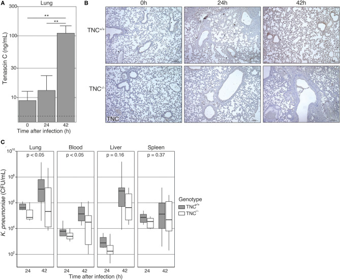
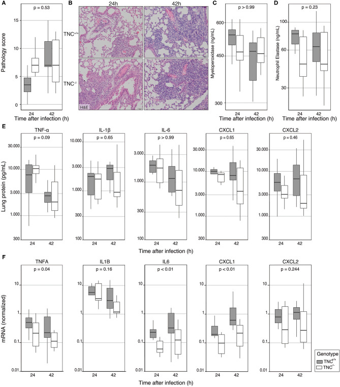
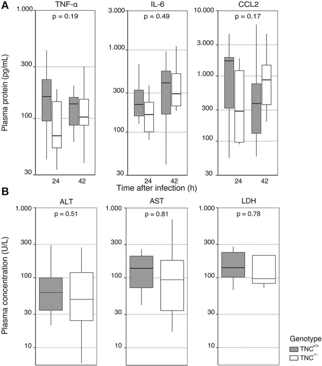
Tenascin C (TNC) is an extracellular matrix glycoprotein that recently emerged as an immunomodulator. TNC-deficient (TNC-/-) mice were reported to have a reduced inflammatory response upon systemic administration of lipopolysaccharide, the toxic component of gram-negative bacteria. Here, we investigated the role of TNC during gram-negative pneumonia derived sepsis. TNC+/+ and TNC-/- mice were infected with Klebsiella pneumoniae via the airways and sacrificed 24 and 42 h thereafter for further analysis. Pulmonary TNC protein levels were elevated 42 h after infection in TNC+/+ mice and remained undetectable in TNC-/- mice. TNC-/- mice showed modestly lower bacterial loads in lungs and blood, and a somewhat reduced local-but not systemic-inflammatory response. Moreover, TNC-/- and TNC+/+ mice did not differ with regard to neutrophil recruitment, lung pathology or plasma markers of distal organ injury. These results suggest that while TNC shapes the immune response during lipopolysaccharide-induced inflammation, this role may be superseded during pneumosepsis caused by a common gram-negative pathogen.

Keywords: Klebsiella pneumoniae (K. pneumoniae); alarmins; immune system; innate immunity; mice; pneumonia; sepsis; tenascin C.


Conflict of interest statement

The authors declare that the research was conducted in the absence of any commercial or financial relationships that could be construed as a potential conflict of interest.

Figures

Figure 1
Figure 1
Pulmonary Tenascin-C levels and bacterial counts during Klebsiella induced pneumosepsis. Tenascin C sufficient (TNC+/+) and deficient (TNC−/−) mice were intranasally infected with K. pneumoniae. (A) Before inoculation, and 24 and 42 h thereafter lung samples were collected and homogenized. TNC was measured in the lysate of 4 TNC+/+ mice from each time point. Bars and whiskers show mean and SE. The dotted line represents the lower limit of quantitation. TNC was not detectable in any of the TNC−/− samples measured. (B) Lung tissue was collected, fixed and embedded in paraffin 24 and 42 h after infection, as well as in the naïve state. Tissue slides were stained for TNC protein. Depicted slides are representative of 5 independent biological replicates. (C) Number of colony-forming units (CFU) 24 and 42 h after infection. Data are shown as Tukey boxplots without outliers. **p < 0.01 in an unpaired t-test. P-values (C) represent the effect of genotype across time-points, as indicated by a two-way type III ANOVA.
Figure 2
Figure 2
Endogenous Tenascin-C does not impact lung inflammation during Klebsiella induced pneumosepsis. TNC+/+ and TNC−/− mice were intranasally infected with K. pneumoniae; 24 and 42 h after infection, lung tissue samples were collected for histological examination or homogenization. (A) Lung samples of all mice were scored for interstitial inflammation, edema, endothelialitis, bronchitis, pleuritis, and the presence of thrombi by a blinded pathologist blinded for the group identity, after which the total pathology score was calculated. (B) Representative pictures of lung pathology at 24 h (middle panel) and 42 hours (right panel) after infection in TNC+/+ mice (top panel) and TNC−/− mice (bottom panel). (C) Levels of MPO, (D) elastase, and (E) cytokines and chemokines were measured in lung homogenates. (F) Cytokine mRNA was measured in RNA isolated from lung homogenate, and is presented normalized to HPRT1 expression. Data are shown as Tukey boxplots without outliers. p-values represent the effect of genotype across time-points, as indicated by a two-way type III ANOVA.
Figure 3
Figure 3
Endogenous Tenascin-C does not impact systemic inflammation during Klebsiella induced pneumosepsis. TNC+/+ and deficient TNC−/− mice were infected with K. pneumoniae; (A) 24 and 42 h after infection plasma was collected for measurements of TNF-α, IL-6, and CCL2. (B) Markers of hepatic injury (ALT, AST) and cell injury in general (LDH) were measured at 42 h. Data are shown as Tukey boxplots without outliers. p-values represent the effect of genotype across time-points, as indicated by a two-way type III ANOVA (A), or unpaired t-test (B).

Similar articles

Cited by

References

    1. Midwood KS, Chiquet M, Tucker RP, Orend G. Tenascin-C at a glance. J Cell Sci. (2016) 129:4321–7. 10.1242/jcs.190546 - DOI - PubMed
    1. Marzeda AM, Midwood KS. Internal affairs: tenascin-c as a clinically relevant, endogenous driver of innate immunity. J Histochem Cytochem. (2018) 66:289–304. 10.1369/0022155418757443 - DOI - PMC - PubMed
    1. Midwood K, Sacre S, Piccinini AM, Inglis J, Trebaul A, Chan E, et al. . Tenascin-C is an endogenous activator of Toll-like receptor 4 that is essential for maintaining inflammation in arthritic joint disease. Nat Med. (2009) 15:774–80. 10.1038/nm.1987 - DOI - PubMed
    1. Goh FG, Piccinini AM, Krausgruber T, Udalova IA, Midwood KS. Transcriptional regulation of the endogenous danger signal tenascin-C: a novel autocrine loop in inflammation. J Immunol. (2010) 184:2655–62. 10.4049/jimmunol.0903359 - DOI - PubMed
    1. Deligne C, Murdamoothoo D, Gammage AN, Gschwandtner M, Erne W, Loustau T, et al. . Matrix-targeting immunotherapy controls tumor growth and spread by switching macrophage phenotype. Cancer Immunol Res. (2020) 8:368–82. 10.1158/2326-6066.Cir-19-0276 - DOI - PMC - PubMed

Publication types