Review
. 2021 Apr;16(2):99-108.
doi: 10.1016/j.joto.2020.09.002. Epub 2020 Sep 10.

The translabyrinthine approach in residency training. "Step by step" surgical anatomy from the dissection laboratory to the operative room

Affiliations
Review

The translabyrinthine approach in residency training. "Step by step" surgical anatomy from the dissection laboratory to the operative room

Fabio Roberti et al. J Otol. 2021 Apr.

Abstract

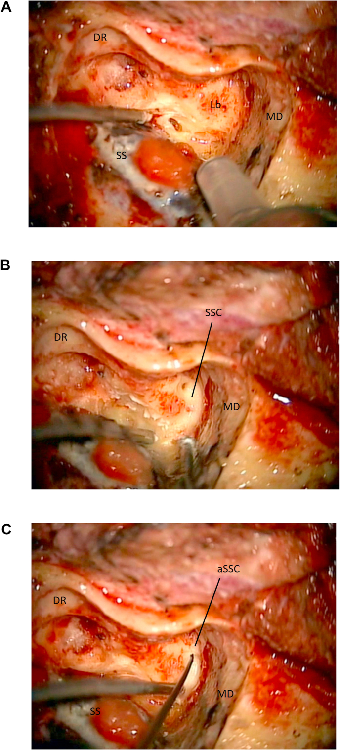
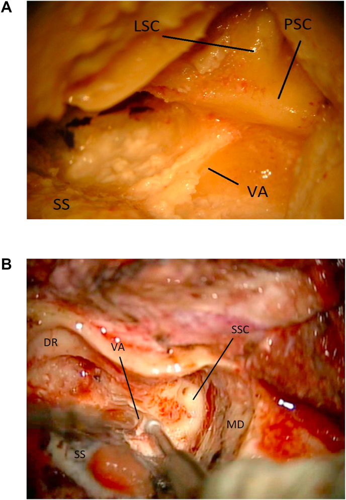
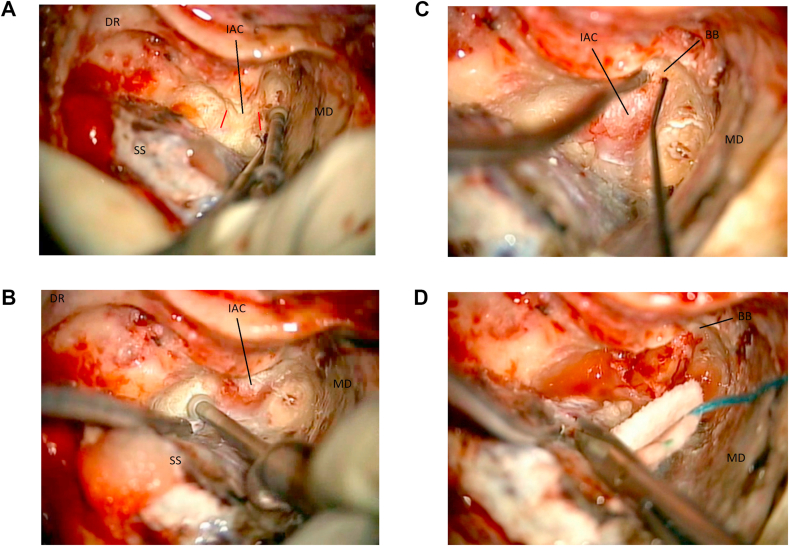
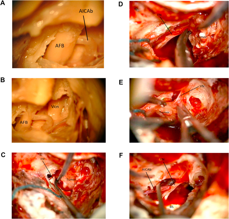
Surgical anatomy training in a dedicated research laboratory and attendance to focused "hands-on" dissection courses are of high educational importance in order to acquire and maintain surgical expertise in skull base surgery, both for young and more experienced surgeons. Nevertheless, transitioning surgical skills and anatomic knowledge from the laboratory to the operative room it is not free of challenges, especially during skull base approaches where the three-dimensional surgical orientation can be quite complex. We present a "step-by-step" and "side-by-side" surgical anatomy report on a translabyrinthine approach that was practiced in the laboratory then performed in the operative room by the surgical team, and we compare surgical anatomy exposures while discussing intraoperative techniques, nuances and challenges, both in the laboratory and the operative room.

Keywords: EMG, electromyogram; OR, operative room; Skull base surgery; TL, translabyrinthine; Translabyrinthine approach; Vestibular schwannomas.


Figures

Fig. 1
Fig. 1
Left TL, supine position with head rotated. Skin incision is marked. 1a: (Lab) periosteum is dissected away from the mastoid that is now exposed. Spine of Henle is visualized. 1 b:(OR) mastoid, posterior temporal and suboccipital regions are exposed, periosteum has been dissected away.
Fig. 1
Fig. 1
Left TL, supine position with head rotated. Skin incision is marked. 1a: (Lab) periosteum is dissected away from the mastoid that is now exposed. Spine of Henle is visualized. 1 b:(OR) mastoid, posterior temporal and suboccipital regions are exposed, periosteum has been dissected away.
Fig. 2
Fig. 2
2a: (Lab) “Approach Triangle” has been outlined. 2 b:(OR) Mastoidectomy within the “Approach Triangle” is started.
Fig. 3
Fig. 3
3a: (Lab) Sigmoid sinus (SS) is located and the antrum (An) is opened. 3 b: (OR) The antrum (An) is opened.
Fig. 4
Fig. 4
4a: (Lab) The short process of the incus (In) is visualized. The sigmoid sinus (SS) is marked. 4 b: (OR) The aditus ad antrum has been opened and the short process of the incus (In) is found. Sigmoid sinus (SS) is marked for orientation.
Fig. 5
Fig. 5
5a: (Lab) The digastric ridge (DR) is located. The course of the facial nerve (FN), the sigmoid sinus (SS) and the bony labyrinth (Lb) are marked. 5 b: (OR) The digastric ridge (DR) is located in front of the sigmoid sinus (SS). Middle fossa dura (MD) is exposed.
Fig. 6
Fig. 6
6a: (Lab) Laboratory image of the exposed anatomy once the mastoidectomy is completed. The labyrinth is visualized. Location of the subarcuate artery is shown (SA). Superior semicircular canal (SSC). Lateral semicircular canal (LSC). Posterior semicircular canal (PSC). Sino-dural angle (SDA). Incus (In). Middle fossa dura (MD). Vestibular Aqueduct (VA). Sigmoid sinus (SS). 6 b: (OR). The arcuate eminence (AE) is located by gently elevating the middle fossa dura (MD). Sigmoid sinus (SS). 6c: (OR) The sino-dural angle (SDA) is exposed. Sigmoid sinus (SS). Middle fossa dura (MD).
Fig. 7
Fig. 7
(Lab) The jugular bulb (JB) is exposed. Labyrinth (Lb). Middle fossa dura (MD). Sigmoid sinus (SS). Facial nerve (FN). Digastric ridge (DR).
Fig. 8
Fig. 8
8a: (OR) The labyrinth (Lb) is located at the end of the mastoidectomy. Middle fossa dura (MD). Sigmoid sinus (SS). Digastric ridge (DR). 8 b: (OR) The labyrinthectomy is carried out. Here the superior semicircular (SSC) and its membranous labyrinth are opened. Middle fossa dura (MD). 8c: (OR) The ampulla of the superior semicircular canal (aSSC) is opened and kept as landmark for the fundus. Middle fossa dura (MD). Sigmoid sinus (SS). Digastric ridge (DR).
Fig. 9
Fig. 9
9a: (Lab) The vestibular aqueduct/endolymphatic sac (VA) are marked after a partial labyrinthectomy. Sigmoid sinus (SS). Lateral semicircular canal (LSC). Posterior semicircular canal (PSC). 9 b: (OR) The vestibular aqueduct (VA) is unroofed while completing the labyrinthectomy. Middle fossa dura (MD). Sigmoid sinus (SS). Digastric ridge (DR). Superior semicircular canal (SSC).
Fig. 10
Fig. 10
10a: (OR) The dura of the internal acoustic canal (IAC) is skeletonized. The orientation of the canal is outlined in red. Middle fossa dura (MD). Sigmoid sinus (SS). Digastric ridge (DR). 10 b: (OR) The internal acoustic canal (IAC) is exposed. Middle fossa dura (MD). Sigmoid sinus (SS). Digastric ridge (DR). 10c: (OR) lateral exposure of the internal acoustic canal (IAC) with direct stimulation (nerve stimulator is seen in the field). The vertical crest/Bill’s bar (BB) is located. Middle fossa dura (MD). 10 d: (OR) The posterior fossa dura is opened and the end of the TL approach. Middle fossa dura (MD). Bill bar (BB).
Fig. 11
Fig. 11
11a: (Lab) Dissection images of the acoustic-facial bundle (AFB) within the IAC. A branch of the Anterior inferior cerebellar artery is seen (AICAb). 11 b: (Lab) Dissection image after removal of portion of the middle fossa dura. Acoustic-facial bundle (AFB). Trigeminal nerve (Vcn). 11c: (OR) Intraoperative image. A vestibular schwannoma (Tumor) is being dissected away from the facial nerve (FN) near the brainstem. 11 d: (OR) Intraoperative image. The vestibular schwannoma (Tumor) is being dissected away from the facial nerve (FN) more laterally. 11e: (OR) Intraoperative image. The vestibular schwannoma (Tumor) is being dissected away from the vestibular nerve (VN). 11f: (OR) Intraoperative image. The tumor has been removed. Brainstem (BS). Anterior inferior cerebellar artery branch (AICAb). Facial nerve (FN).
Fig. 12
Fig. 12
12a: (OR) The incus (In) and the ossicles are removed from the middle ear before final closure. 12 b: (OR) The middle ear is gently packed with Surgicel to avoid postoperative CSF leak. Sigmoid sinus (SS). Digastric ridge (DR). 12c: (OR) Abdominal fat graft and fibrin glue are utilized to fill the mastoidectomy cavity before myocutaneous closure.

References

    1. Ammar M.B., Piccirillo E., Topsakal V., Taibah A., Sanna M. Surgical results and technical refinements in translabyrinthine excision of vestibular schwannomas: the gruppo otologico experience. Neurosurgery. Jun 2012;70(6):1481–1491. - PubMed
    1. Cushing H. WB Saunders Co; Philadelphia, PA: 1917. Tumors of the Nervus Acusticus and the Syndrome of the Cerebellopontine Angle.
    1. Cushing H. Further concerning the acoustic neuromas. Laryngoscope. 1921;31:209–228.
    1. Hirsch B.E., Cass S.P., Sekhar L.N., Wright D.C. Translabyrinthine approach to skull base tumors with hearing preservation. Am. J. Otol. 1993;14:533–543. - PubMed
    1. House W.F. Transtemporal bone microsurgical removal of acoustic neuromas. Report of cases. Arch. Otolaryngol. 1964;80:617–667. - PubMed

LinkOut - more resources