

doi: 10.1155/2021/6661828.
eCollection 2021.
miR-22-3p/PGC1 β Suppresses Breast Cancer Cell Tumorigenesis via PPAR γ
Affiliations
- PMID: 33777130
- PMCID: PMC7981180
- DOI: 10.1155/2021/6661828
Item in Clipboard
miR-22-3p/PGC1 β Suppresses Breast Cancer Cell Tumorigenesis via PPAR γ
PPAR Res.
.
Abstract
In this study, we found that miR-22-3p expression was decreased in breast cancer (BC) cell lines and tissues. Overexpression of miR-22-3p inhibited the proliferation and migration of BC cells in vitro and in vivo, while depletion of miR-22-3p exhibited the opposite effect. Importantly, miR-22-3p could directly target PGC1β and finally regulate the PPARγ pathway in BC. In conclusion, miR-22-3p/PGC1β suppresses BC cell tumorigenesis via PPARγ, which may become a potential biomarker and therapeutic target.
Copyright © 2021 Xuehui Wang et al.
Conflict of interest statement
The authors declare that they have no competing interests.
Figures
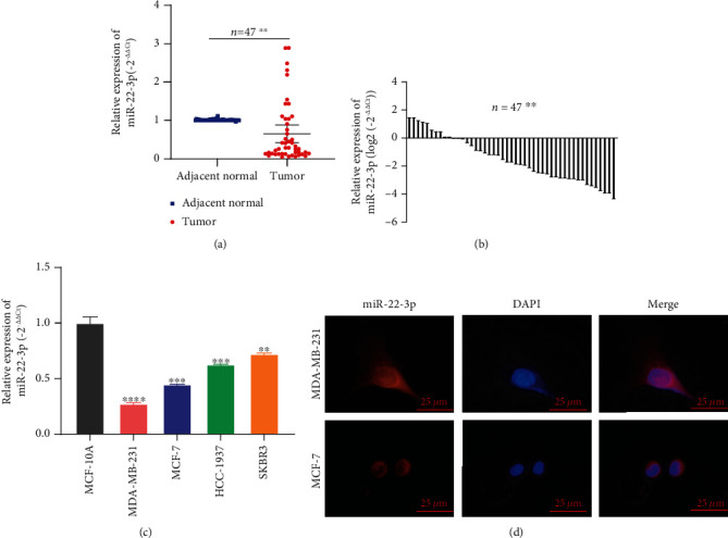
miR-22-3p was decreased in BC cell lines and tissues. (a, b) miR-22-3p had low expression in BC tissues compared with adjacent normal tissues. (c) miR-22-3p had low expression in BC cell lines. (d) Detection of colocalization of miR-22-3p in cytoplasm by RNA FISH assay (magnification, ×400). Red, miR-200a-3p; blue, DAPI. ∗∗p < 0.1; ∗∗∗p < 0.001; ∗∗∗∗p < 0.0001.
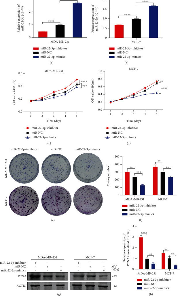
miR-22-3p suppressed cell proliferation of BC cells. (a, b) Expression of miR-22-3p was confirmed by RT-qPCR in MDA-MB-231 and MCF-7 cells. (c, d) Effect of miR-22-3p on proliferation in MDA-MB-231 and MCF-7 cells by MTT assay. (e, f) Effect of miR-22-3p on proliferation in MDA-MB-231 and MCF-7 cells by colony formation assay. (g, h) Effect of miR-22-3p on proliferation in MDA-MB-231 and MCF-7 cells by western blotting. ∗∗p < 0.01; ∗∗∗p < 0.001; ∗∗∗∗p < 0.0001.
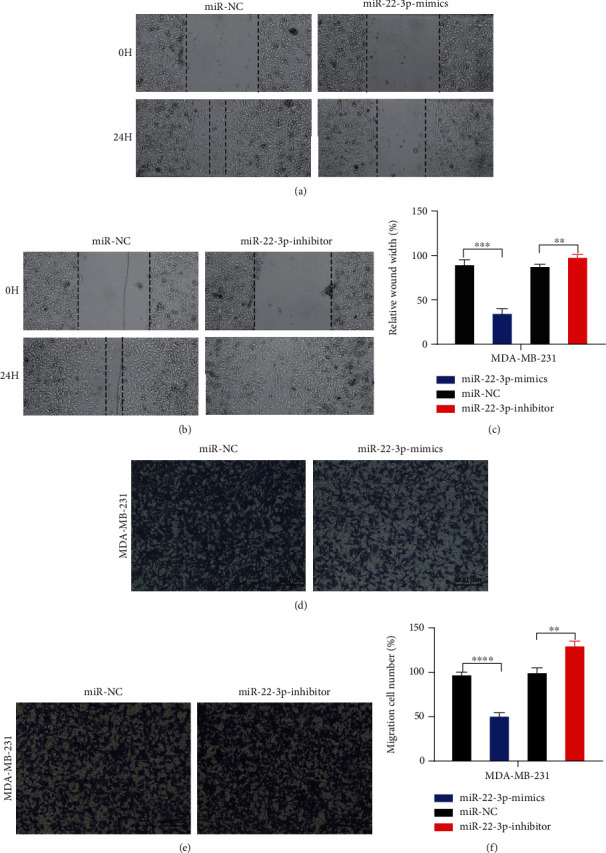
miR-22-3p suppressed cell migration of BC cells. (a–c) Wound healing assays were performed in MDA-MB-231 cell line treated with miR-22-3p mimics or miR-22-3p inhibitor (miR-NC as negative control). (d–f) Cell migration assays were performed in MDA-MB-231 cell line treated with miR-22-3p mimics or miR-22-3p inhibitor (miR-NC as negative control). ∗∗p < 0.01; ∗∗∗p < 0.001; ∗∗∗∗p < 0.0001.
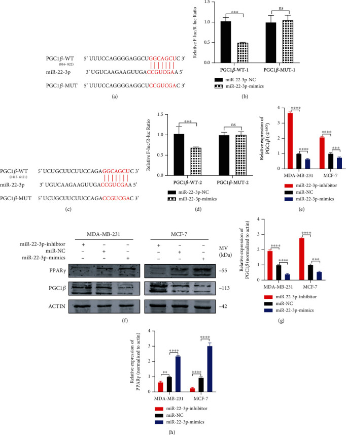
PGC1β is a direct target of miR-22-3p. (a, c) Putative complementary sites within miR-22-3p and PGC1β predicted by bioinformatics analysis (TargetScan). (b, d) Dual-luciferase reporter assays demonstrated that PGC1β is a direct target of miR-22-3p. (e) PGC1β mRNA level was determined by RT-PCR in MDA-MB-231 and MCF-7 cells with different treatment. (f–h) Representative western blots and quantification of PGC1β and PPARγ in MDA-MB-231 and MCF-7 cells with different treatment. β-Actin was used as an internal control. ∗∗p < 0.01; ∗∗∗p < 0.001; ∗∗∗∗p < 0.0001.
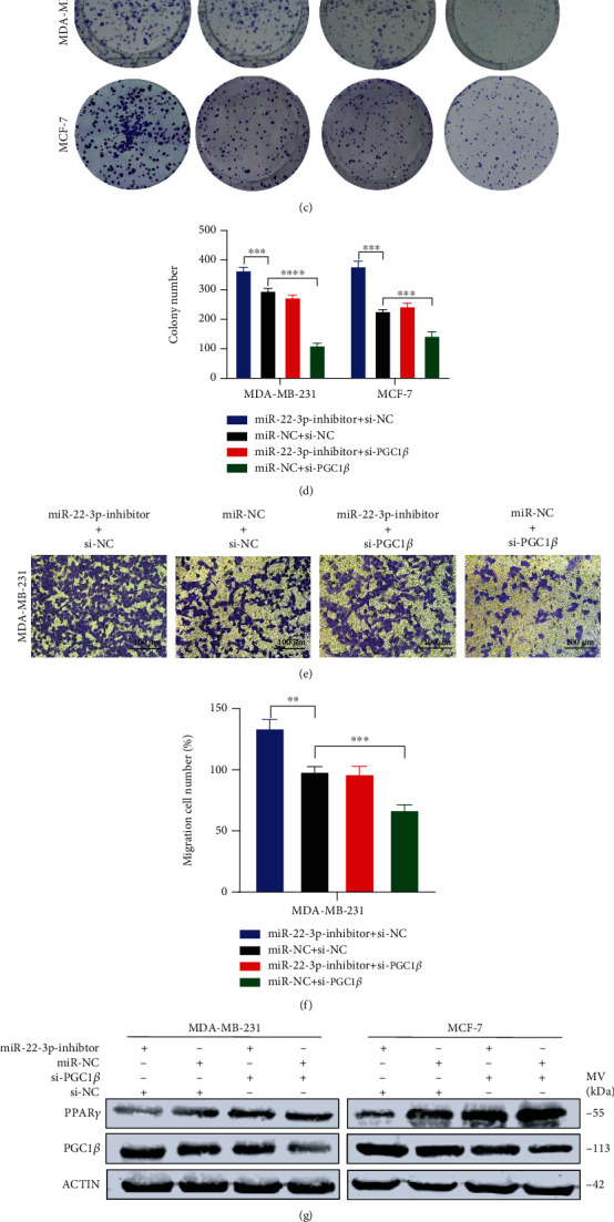
miR-22-3p suppressed the proliferation and migration of BC cells via PGC1β. (a–d) Knockdown of PGC1β partially reversed miR-22-3p inhibitor-induced promotion of proliferation in MDA-MB-231 and MCF-7 cells determined by MTT assay and colony assay. (e, f) Knockdown of PGC1β partially reversed miR-22-3p inhibitor-induced promotion of migration in MDA-MB-231 and MCF-7 cells determined by transwell assay. (g–i) Western blotting analysis for PGC1β/PPARγ protein level in MDA-MB-231 and MCF-7 cells. ∗p < 0.05; ∗∗p < 0.01; ∗∗∗p < 0.001; ∗∗∗∗p < 0.0001.
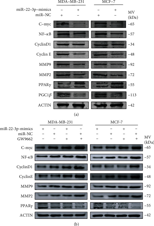
Inhibition of PPARγ attenuates suppression of miR-22-3p on BC cells. (a) Upregulated miR-22-3p increased the expression of PPARγ and decreased the expression of PGC1β, C-myc, NF-κB, CyclinD1, cyclin E, MMP2, and MMP9. (b) Downregulation of C-myc, NF-κB, CyclinD1, cyclin E, MMP2, and MMP9 induced by miR-22-3p was inverted by PPARγ inhibition (GW9662).
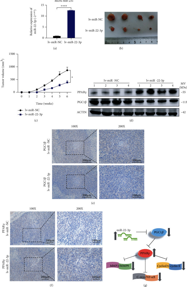
miR-22-3p suppressed BC tumor growth in vivo. (a) Overexpression of miR-22-3p in MDA-MB-231 cells was verified by RT-qPCR. (b) Representative images of xenograft tumors in nude mice. (c) The growth curves of xenografts. (d) Extract protein from tumors and measuring the expression of PGC1β/PPARγ by western blotting. (e, f) Immunohistochemistry (IHC) staining of PGC1β/PPARγ in xenografts. (g) The mechanism diagram was generated to illustrate the mechanism of miR-22-3p-PGC1β-PPARγ in BC. ∗p < 0.05; ∗∗∗∗p < 0.0001.
Similar articles
-
The Role of miR-640: A Potential Suppressor in Breast Cancer via Wnt7b/β-catenin Signaling Pathway.Front Oncol. 2021 Apr 12;11:645682. doi: 10.3389/fonc.2021.645682. eCollection 2021. Front Oncol. 2021. PMID: 33912460 Free PMC article.
-
MiR-3194-3p Inhibits Breast Cancer Progression by Targeting Aquaporin1.Front Oncol. 2020 Aug 7;10:1513. doi: 10.3389/fonc.2020.01513. eCollection 2020. Front Oncol. 2020. PMID: 32903818 Free PMC article.
-
MiR-1301-3p inhibits human breast cancer cell proliferation by regulating cell cycle progression and apoptosis through directly targeting ICT1.Breast Cancer. 2018 Nov;25(6):742-752. doi: 10.1007/s12282-018-0881-5. Epub 2018 Jun 27. Breast Cancer. 2018. PMID: 29951881
-
miR-758-3p suppresses human bladder cancer cell proliferation, migration and invasion by targeting NOTCH2.Exp Ther Med. 2019 May;17(5):4273-4278. doi: 10.3892/etm.2019.7400. Epub 2019 Mar 18. Exp Ther Med. 2019. PMID: 30988799 Free PMC article.
-
A novel long noncoding RNA PGC1β-OT1 regulates adipocyte and osteoblast differentiation through antagonizing miR-148a-3p.Cell Death Differ. 2019 Oct;26(10):2029-2045. doi: 10.1038/s41418-019-0296-7. Epub 2019 Feb 6. Cell Death Differ. 2019. PMID: 30728459 Free PMC article.
Cited by
-
Hsa_circ_0103232 promotes melanoma cells proliferation and invasion via targeting miR-661/RAB3D.Cell Cycle. 2022 Sep;21(17):1811-1826. doi: 10.1080/15384101.2022.2072636. Epub 2022 May 13. Cell Cycle. 2022. PMID: 35549813 Free PMC article.
-
MiR-22-3p suppresses NSCLC cell migration and EMT via targeting RAC1 expression.Funct Integr Genomics. 2023 Aug 25;23(3):281. doi: 10.1007/s10142-023-01211-z. Funct Integr Genomics. 2023. PMID: 37620594 Free PMC article.
-
The Role of miR-1183: A Potential Suppressor in Hepatocellular Carcinoma via Regulating Splicing Factor SRSF1.J Hepatocell Carcinoma. 2023 Jul 21;10:1169-1180. doi: 10.2147/JHC.S408542. eCollection 2023. J Hepatocell Carcinoma. 2023. PMID: 37497429 Free PMC article.
-
Hsa-miR-22-3p inhibits liver cancer cell EMT and cell migration/ invasion by indirectly regulating SPRY2.PLoS One. 2023 Feb 7;18(2):e0281536. doi: 10.1371/journal.pone.0281536. eCollection 2023. PLoS One. 2023. PMID: 36749775 Free PMC article.
-
Development and Validation of a Novel PPAR Signaling Pathway-Related Predictive Model to Predict Prognosis in Breast Cancer.J Immunol Res. 2022 Jun 2;2022:9412119. doi: 10.1155/2022/9412119. eCollection 2022. J Immunol Res. 2022. PMID: 35692496 Free PMC article.
References
LinkOut - more resources
Full Text Sources
Other Literature Sources
