

Laryngeal involvement in a patient with active postprimary tuberculosis: Case report of a rare extrapulmonary manifestation
- PMID: 33777282
- PMCID: PMC7985706
- DOI: 10.1016/j.radcr.2021.02.036
Laryngeal involvement in a patient with active postprimary tuberculosis: Case report of a rare extrapulmonary manifestation
Abstract
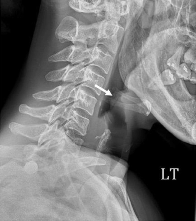
A 43-year-old woman was found to have active post-primary tuberculosis and a lateral neck radiograph showing a thickened epiglottis. Bronchoscopy-guided biopsies of the epiglottis and lung were acid fast bacilli stain positive. Histopathology from both showed multiple caseating granulomas. The patient's condition improved with RIPE therapy. This case illustrates the importance for physicians to be aware of possible laryngeal involvement in tuberculosis and that it can present even without evidence of active or latent tuberculosis.
Keywords: Dysphonia; Extrapulmonary tuberculosis; Laryngitis; Odynophagia; Tuberculosis.
© 2021 The Authors. Published by Elsevier Inc. on behalf of University of Washington.
Figures


Similar articles
-
Laryngeal Tuberculosis in Pregnant Women: A Case Report and Review of the Literature.Cureus. 2018 Nov 5;10(11):e3545. doi: 10.7759/cureus.3545. Cureus. 2018. PMID: 30648077 Free PMC article.
-
Isolated testicular tuberculosis: A case report.Urol Case Rep. 2024 Oct 11;57:102869. doi: 10.1016/j.eucr.2024.102869. eCollection 2024 Nov. Urol Case Rep. 2024. PMID: 39498390 Free PMC article.
-
Unusual Presentation of Extrapulmonary Tuberculosis as Laryngeal Mass in an Atypical Patient.Case Rep Med. 2024 Dec 13;2024:9912317. doi: 10.1155/carm/9912317. eCollection 2024. Case Rep Med. 2024. PMID: 39711754 Free PMC article.
-
Laryngeal tuberculosis.Am J Otolaryngol. 2000 Mar-Apr;21(2):122-6. doi: 10.1016/s0196-0709(00)85010-3. Am J Otolaryngol. 2000. PMID: 10758999 Review.
-
Pathology of postprimary tuberculosis in humans and mice: contradiction of long-held beliefs.Tuberculosis (Edinb). 2007 Jul;87(4):267-78. doi: 10.1016/j.tube.2006.11.003. Epub 2007 Mar 21. Tuberculosis (Edinb). 2007. PMID: 17369095 Review.
References
-
- Uslu C, Oysu C, Uklumen B. Tuberculosis of the epiglottis: a case report. Eur Arch Otorhinolaryngol. 2008;265(5):599–601. - PubMed
-
- Auerbach O. Laryngeal tuberculosis. Arch Otolaryngol. 1946;44:191–201. - PubMed
-
- Rizzo PB, Da mosto MC, Clari M, Scotton PG, Vaglia A, Marchiori C. Laryngeal tuberculosis: an often forgotten diagnosis. Int J Infect Dis. 2003;7(2):129–131. - PubMed
-
- Cleary K.R., Batsakis J.G. Mycobacterial disease of the head and neck: current perspective. Ann Otol Rhinol Laryngol. 1995;104(10 Pt 1):830–833. - PubMed
-
- Bailey C.M., Windle-Taylor P.C. Tuberculous laryngitis: a series of 37 patients. The Laryngoscope. 1981;91(1):93–100. - PubMed
Publication types
LinkOut - more resources
Full Text Sources
Other Literature Sources
