. 2021 Mar 9:2021:1457089.
doi: 10.1155/2021/1457089. eCollection 2021.

Regulation of Superoxide by BAP31 through Its Effect on p22phox and Keap1/Nrf2/HO-1 Signaling Pathway in Microglia

Affiliations

Regulation of Superoxide by BAP31 through Its Effect on p22phox and Keap1/Nrf2/HO-1 Signaling Pathway in Microglia

Xia Liu et al. Oxid Med Cell Longev. .

Abstract

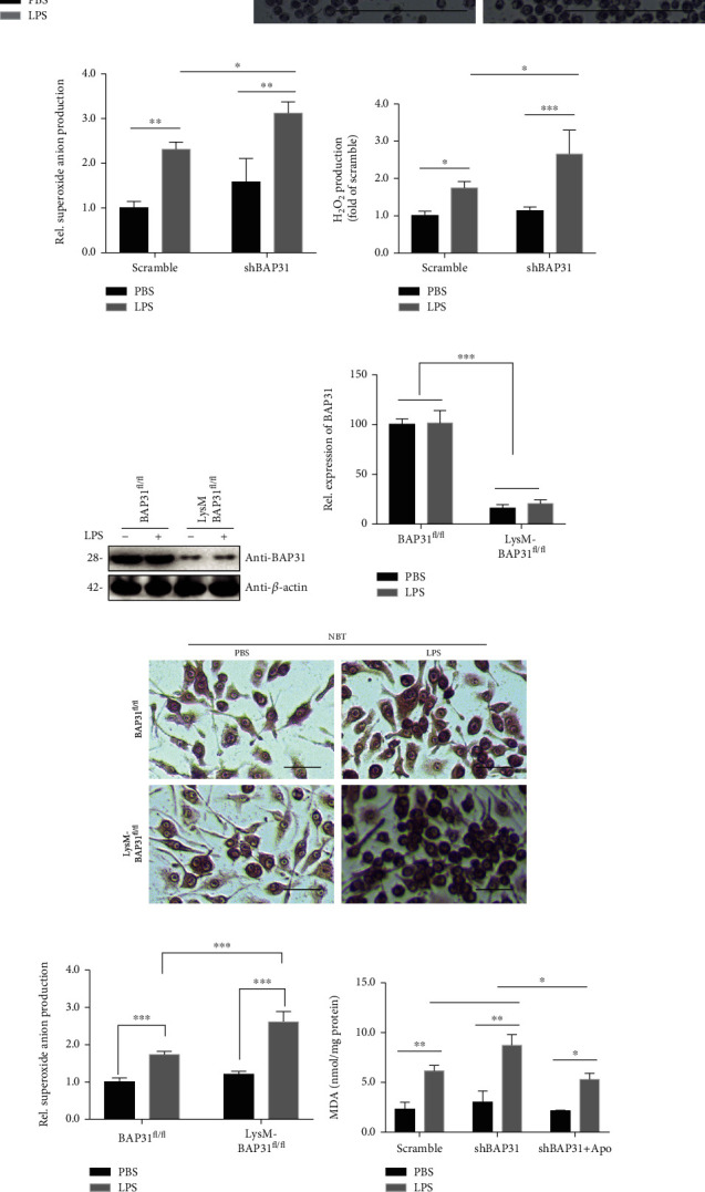
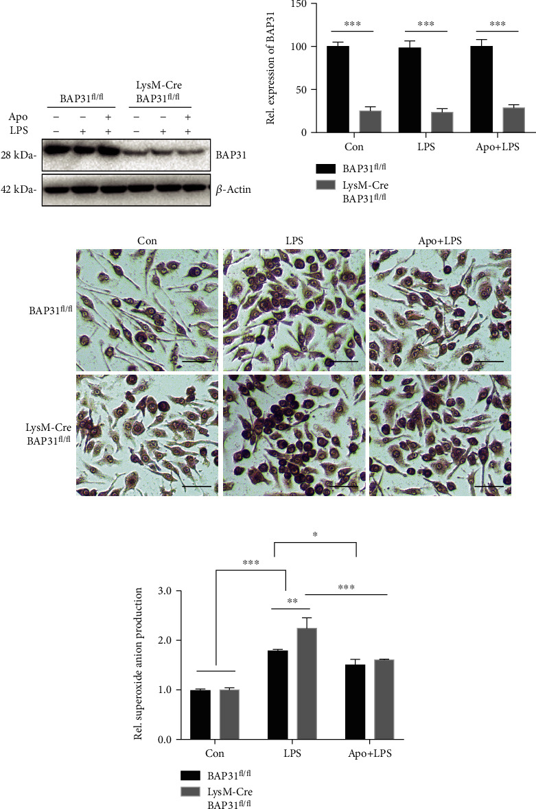
Reactive oxygen species (ROS) production by activation of microglia is considered to be a major cause of neuronal dysfunction, which can lead to damage and death through direct oxidative damage to neuronal macromolecules or derangement of neuronal redox signaling circuits. BAP31, an integral ER membrane protein, has been defined as a regulatory molecule in the CNS. Our latest studies have found that BAP31 deficiency leads to activation of microglia. In this study, we discovered that BAP31 deficiency upregulated LPS-induced superoxide anion production in BV2 cells and mice by upregulating the expression level of p22phox and by inhibiting the activation of Nrf2-HO-1 signaling. Knockdown of p22phox/keap1 or use of an NADPH oxidase inhibitor (apocynin) reversed the production of superoxide anion and inflammatory cytokines, which then reduced neuronal damage and death in vitro and in vivo. These results suggest that BAP31 deficiency contributes to microglia-related superoxide anion production and neuroinflammation through p22phox and keap1. Furthermore, the excess superoxide anion cooperated with inflammatory cytokines to induce the damage and death of neurons. Thus, we determined that BAP31 is an important regulator in superoxide anion production and neuroinflammation, and the downstream regulators or agonists of BAP31 could therefore be considered as potential therapeutic targets in microglial-related superoxide anion production and neuroinflammation.


Conflict of interest statement

The authors declare that they have no competing interests.

Figures

Figure 1
Figure 1
BAP31 deficiency upregulates p22phox and keap1 and inhibits Nrf2/HO-1 signaling in LPS-treated BV2 microglia. Scramble and shBAP31 were treated with LPS (100 ng/ml) for 24 h. The mRNA levels of p22phox (a), p40phox (b), p67phox (c), and gp91phox (d) were analyzed with RT-PCR. (e) Representative Western blots showing the expression levels of keap1 in scramble and shBAP31 BV2 cells. (f) Scramble and shBAP31 cells were treated with LPS for 24 h. The protein levels of Nrf2, BAP31, and β-actin were analyzed by Western blotting. (g) Scramble and shBAP31 BV2 cells were treated with LPS for 24 h; the cytosolic and nuclear fractions of Nrf2 were analyzed by Western blotting with antibodies against Nrf2, histone, and β-actin. (h) Immunoblots for Nrf2 in cytosolic fractions were quantified and normalized to β-actin protein. (i) Immunoblots for Nrf2 in nuclear fractions were quantified and normalized to histone protein. (j) Representative Western blots showing the expression levels of HO-1 in scramble and shBAP31 BV2 cells after LPS treatment for 24 h. All the data are indicated as mean ± SEM of three independent experiments. p < 0.05, ∗∗p < 0.01, and ∗∗∗p < 0.001 versus the control group.
Figure 2
Figure 2
BAP31 deficiency exacerbates LPS-induced superoxide anion generation in microglia. (a) The protein level of BAP31 in scramble and shBAP31 BV2 microglial cells. (b) Scramble and shBAP31 cells were stimulated with LPS (100 ng/ml) for 12 h. Superoxide anion were measured by staining with NBT and observed by confocal microscopy. Scale bars = 100 μm. (c) Intracellular superoxide anion detected by NBT staining in (b) was quantified using a multimode microplate reader. (d) Scramble and shBAP31 cells were stimulated with LPS (100 ng/ml) for 12 h; H2O2 production was measured using a multimode microplate reader. (e, f) The protein level of BAP31 in primary microglial cells from BAP31fl/fl and LysM-Cre-BAP31fl/fl mice. (g) Visualization of NBT staining in primary microglial cells from BAP31fl/fl and LysM-Cre-BAP31fl/fl mice after LPS treatment (100 ng/ml) for 12 h. (h) Intracellular superoxide anion detected by NBT staining in (g) were quantified using a multimode microplate reader. Scale bars = 10 μm. (i) Scramble and shBAP31 cells were pretreated with apocynin for 1 h and then stimulated with LPS (100 ng/ml) for 24 h. Lipid peroxidation (MDA) levels were measured using a multimode microplate reader. Data are indicated as the mean ± SEM of three independent experiments. p < 0.05, ∗∗p < 0.01, and ∗∗∗p < 0.001 versus the control group.
Figure 3
Figure 3
BAP31 deficiency induces proinflammatory cytokine production in LPS-stimulated cells. Scramble and shBAP31 BV2 cells were treated with LPS (100 ng/ml) for 24 h. The mRNA levels of BAP31 (a), IL-1β (b), TNFα (c), CD80 (d), CD86 (e), Arg1 (f), CD206 (g), Fizz1 (h), and Ym1 (i) were analyzed by RT-PCR. Flow cytometry shows that the expression of CD86 (j) and CD206 (k) in primary microglial cells were treated with LPS (100 ng/ml) for 24 h. All the data are indicated as mean ± SEM of three independent experiments. p < 0.05, ∗∗p < 0.01, and ∗∗∗p < 0.001 versus the control group.
Figure 4
Figure 4
Knockdown of p22phox or keap1 prevents BAP31-deficiency-induced superoxide anion production upon LPS stimulation. (a, b) Primary microglial cells were transfected with p22phox siRNA for 60 h, followed by treatment with LPS for 12 h. The relative superoxide anion levels were measured by staining with NBT and quantified using a multimode microplate reader. (c, d, e) Primary microglial cells were transfected with keap1 siRNA for 60 h, followed by treatment with LPS for 24 h. The cytosolic and nuclear fractions were analyzed by Western blotting with antibodies against Nrf2, histone, and β-actin. (f) Primary microglial cells were transfected with keap1 siRNA for 60 h, followed by treatment with LPS for 12 h. The relative superoxide anion was measured by staining with NBT and quantified using a multimode microplate reader. All the data are indicated as mean ± SEM of three independent experiments. p < 0.05, ∗∗p < 0.01, and ∗∗∗p < 0.001 versus the control group.
Figure 5
Figure 5
Apocynin alleviates superoxide anion production caused by the deficiency of BAP31. (a, b) Protein levels of BAP31 in primary microglial cells from BAP31fl/fl and LysM-Cre-BAP31fl/fl mice. (c, d) Visualization of NBT staining in primary microglial cells from BAP31fl/fl and LysM-Cre-BAP31fl/fl mice pretreated with apocynin for 1 h and then stimulated with LPS (100 ng/ml) for 12 h. Intracellular superoxide anion detected by NBT staining were quantified using a multimode microplate reader. Scale bars = 10 μm. All the data are indicated as mean ± SEM of three independent experiments. p < 0.05, ∗∗p < 0.01, and ∗∗∗p < 0.001 versus the control group.
Figure 6
Figure 6
Knockdown of p22phox or keap1 prevents proinflammatory cytokine production in BV2 microglia. Scramble and shBAP31 were transfected with p22phox or keap1 siRNA for 60 h, followed by treatment with LPS for 24 h. The mRNA levels of BAP31 (a), p22phox (b), keap1 (c), IL-1β (d), TNFα (e), CD80 (f), and CD86 (g) were analyzed by RT-PCR. All the data are indicated as mean ± SEM of three independent experiments. p < 0.05, ∗∗p < 0.01, and ∗∗∗p < 0.001 versus the control group.
Figure 7
Figure 7
Apocynin prevents proinflammatory cytokine release in BV2 microglia and alleviates LPS-induced neurotoxicity among cocultures. (a, b, and c) Scramble and shBAP31 BV2 cells were treated with LPS (100 ng/ml) for 24 h, and the secreted protein levels of the cytokines IL-1β (a) and TNFα (b) in the supernatant were analyzed using ELISA kits. NO production (c) was measured by the Griess assay. (d, e) Visualization of SHSY5Y cells after coculture with microglial conditional medium (MCM) from scramble and shBAP31 BV2 cells exposed to LPS for 24 h after treatment with apocynin for 1 h. Cell viability was measured by the MTT assay. Scale bars = 200 μm. All the data are indicated as mean ± SEM of three independent experiments. p < 0.05, ∗∗p < 0.01, and ∗∗∗p < 0.001 versus the control group.
Figure 8
Figure 8
Apocynin prevents BAP31-deficiency-induced superoxide anion production in vivo. (a) Representative images of DHE staining from the hippocampus and cortex after LPS administration. Quantification of DHE staining of the hippocampus (b) and cortex (c) in BAP31fl/fl and LysM-Cre-BAP31fl/fl mice. Scale bars = 200 μm. n = 8 per group for each experiment. Data are expressed as mean ± SEM. p < 0.05, ∗∗p < 0.01, and ∗∗∗p < 0.001 versus the control group.
Figure 9
Figure 9
Conditional microglial BAP31 knockout mice exhibit more inflammation and oxidative damage when administered LPS. (a) A representative schematic diagram and microscopy images of 8-OHdG (green), NeuN (red), and DAPI (blue) triple immunofluorescent staining of the brain. Scale bars = 200 μm. (b) Quantification of 8-OHdG (green) and NeuN (red) double positive cells. (c, d) Levels of IL-1β and TNFα mRNA in samples of the hippocampus were analyzed by RT-PCR. All the data are indicated as mean ± SEM of three independent experiments. p < 0.05, ∗∗p < 0.01, and ∗∗∗p < 0.001 versus the control group.
Figure 10
Figure 10
Apocynin protects hippocampal neurons from BAP31-deficiency-induced superoxide anion and inflammation in vivo. (a) Representative images of NeuN-labeled intact neurons in the hippocampal DG areas. The intact neuron is shown in green. Scale bars = 200 μm. (b) Relative fluorescence intensity of NeuN+ cells in the DG. n = 8 per group in each experiment. Data are expressed as mean ± SEM. p < 0.05, ∗∗p < 0.01, and ∗∗∗p < 0.001 versus the control group.
Figure 11
Figure 11
A schematic illustration of BAP31 regulating superoxide anion production and neuroinflammation in microglia. BAP31 deficiency upregulates LPS-induced superoxide anion and hydrogen peroxide production through p22phox and the keap1/Nrf2/HO-1 signaling pathway, excess superoxide anion, and hydrogen peroxide cooperate with inflammatory cytokine to induce the damage and death of neurons.

Similar articles

Cited by

References

    1. Paulsen J. S., Nance M., Kim J.-I., et al. A review of quality of life after predictive testing for and earlier identification of neurodegenerative diseases. Progress in Neurobiology. 2013;110:2–28. doi: 10.1016/j.pneurobio.2013.08.003. - DOI - PMC - PubMed
    1. Rotermund C., Machetanz G., Fitzgerald J. C. The therapeutic potential of metformin in neurodegenerative diseases. Frontiers in Endocrinology. 2018;9:p. 400. doi: 10.3389/fendo.2018.00400. - DOI - PMC - PubMed
    1. Brennan S., Keon M., Liu B., Su Z., Saksena N. K. Panoramic visualization of circulating microRNAs across neurodegenerative diseases in humans. Molecular Neurobiology. 2019;56(11):7380–7407. doi: 10.1007/s12035-019-1615-1. - DOI - PMC - PubMed
    1. Zeng J., Dong Z., Wu H., Tian Z., Zhao Z. Redox regulation of plant stem cell fate. The EMBO Journal. 2017;36(19):2844–2855. doi: 10.15252/embj.201695955. - DOI - PMC - PubMed
    1. Han B. H., Zhou M. L., Johnson A. W., et al. Contribution of reactive oxygen species to cerebral amyloid angiopathy, vasomotor dysfunction, and microhemorrhage in aged Tg2576 mice. Proceedings of the National Academy of Sciences of the United States of America. 2015;112(8):E881–E890. doi: 10.1073/pnas.1414930112. - DOI - PMC - PubMed

MeSH terms

LinkOut - more resources