. 2021 Feb;82(1):96-99.
doi: 10.1055/s-0040-1722636. Epub 2021 Feb 2.

Not a Tumor-Nonspecific Orbital Inflammation

Affiliations

Not a Tumor-Nonspecific Orbital Inflammation

James A Garrity. J Neurol Surg B Skull Base. 2021 Feb.

Abstract

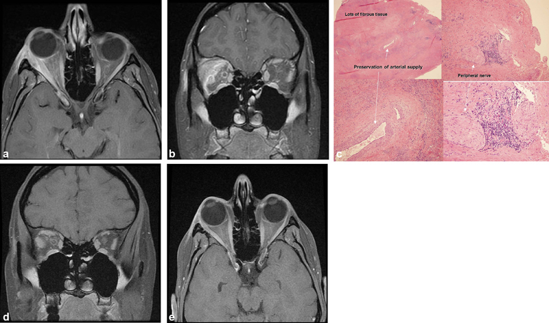
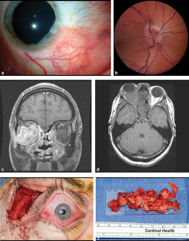
Objective This study was aimed to illustrate the features and complexities of nonspecific orbital inflammation via discussion of two representative cases. Design Present study is a retrospective case review. Setting The study was conducted at a tertiary care medical center. Participants Two patients with nonspecific orbital inflammation were participants of this retrospective study. Main Outcome Measures Outcome of the study was disease-free patients and off all medications. Results At follow-up, both patients are disease free and off all medications. Conclusion Surgery plays a diagnostic and therapeutic role. While the clinical subtype is important for differential diagnosis and symptomatic treatment, the histologic subtype is similarly important. For inflammatory dacryoadenitis, surgery can be therapeutic. For extensive granulomatosis with polyangiitis, debulking surgery may allow better penetration of medications, especially rituximab.

Keywords: dacryoadenitis; granulomatosis with polyangiitis; inflammation; orbit; orbital pseudotumor.


Conflict of interest statement

Conflict of Interest None declared.

Figures

Fig. 1
Fig. 1
( A, B ) Axial and coronal magnetic resonance imaging (MRI), T1 with contrast: the right lacrimal gland and lateral rectus are enlarged and enhance as do the lateral/superior periorbita and optic nerve sheath; stranding of the adjacent orbital fat. ( B ) Coronal MRI, T1 with contrast: the right lacrimal gland and lateral rectus are enlarged and enhance as does the lateral/superior periorbita and the optic nerve sheath. There is also stranding of the adjacent orbital fat. ( C ) Right lacrimal gland. Hematoxylin and eosin (H&E) × 20 upper left, × 100 lower left and upper right, ×200 lower right: note the abundance of fibrous tissue and loss of lacrimal gland architecture with preservation of arterial supply. There is a peripheral nerve with adjacent inflammation which may account for some of the discomfort associated with this condition. ( D, E ) Axial and coronal MRI, T1 with contrast: postoperative scan at 6 months shows mild residual enlargement of right superior and lateral rectus muscles and resection of lacrimal gland mass.
Fig. 2
Fig. 2
( A ) Slit lamp photograph of right cornea showing a pseudopterygium characteristic of healed marginal keratitis from 3:00 through 6:00. ( B ) Fundus photograph of right eye demonstrates swollen optic disk. This resolved following debulking surgery. ( C ) Axial magnetic resonance imaging (MRI) T1 without contrast shows that the right orbit is virtually filled with fibrous tissue. ( D ) Coronal MRI, T1 with contrast: the right medial, inferior and lateral rectus muscles are mildly enlarged and a contrast enhancing infiltrate surrounds these muscles and the optic nerve. There is a small infiltrate in the medial portion of the left orbit. ( E ) Intraoperative photography of a no-bone flap lateral orbitotomy with sutures around each rectus muscle insertion. Gentle traction on suture is transmitted to corresponding muscle belly allowing identification of each encased muscle. All of the palpable retrobulbar tissue was removed through this incision. ( F ) Gross pathology of debulked orbital fibrous tissue.

Similar articles

Cited by

References

    1. Birch-Hirschfeld A. 2nd ed. Berlin, Germany: Springer-Verlag; 1930. Handbuch der Gesamten Augenheilkunden: Die Krankheiten der Orbita.
    1. Jakobiec F A, Font R L. Philadelphia, PA: W.B. Saunders; 1986. Orbit; pp. 2777–2795.
    1. Nugent R A, Rootman J, Robertson W D, Lapointe J S, Harrison P B. Acute orbital pseudotumors: classification and CT features. AJR Am J Roentgenol. 1981;137(05):957–962. - PubMed
    1. Kennerdell J S, Dresner S C. The nonspecific orbital inflammatory syndromes. Surv Ophthalmol. 1984;29(02):93–103. - PubMed
    1. Mahr M A, Salomao D R, Garrity J A. Inflammatory orbital pseudotumor with extension beyond the orbit. Am J Ophthalmol. 2004;138(03):396–400. - PubMed