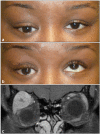
Masses of the Lacrimal Gland: Evaluation and Treatment
- PMID: 33777623
- PMCID: PMC7987400
- DOI: 10.1055/s-0040-1722700
Masses of the Lacrimal Gland: Evaluation and Treatment
Abstract
Lacrimal gland lesions account for approximately 9 to 10% of all biopsied orbital masses. Potential causes include nongranulomatous and granulomatous inflammation, autoimmune disease, lymphoproliferative disorders, benign epithelial proliferation, malignant neoplasia, and metastatic disease. Inflammatory lesions and lymphoproliferative disorders are the most common and may be unilateral or bilateral; they may also be localized to the orbit or associated with systemic disease. Both benign and malignant epithelial lacrimal gland masses tend to be unilateral and involve the orbital lobe, but a more rapid onset of symptoms and periorbital pain strongly suggest malignant disease. On orbital imaging, both inflammatory and lymphoproliferative lesions conform to the globe and surrounding structures, without changes in adjacent bone, whereas epithelial lacrimal gland masses often show scalloping of the lacrimal gland fossa. Malignant epithelial lacrimal gland tumors can also have radiographic evidence of bony invasion and destruction. Masses of the lacrimal gland may be due to a broad range of pathologies, and a good working knowledge of common clinical characteristics and radiographic imaging findings is essential for diagnosis and treatment. All patients with inflammatory, lymphoproliferative, and epithelial neoplastic lesions involving the lacrimal gland require long-term surveillance for disease recurrence and progression.
Keywords: adenoid cystic carcinoma; carcinoma ex pleomorphic adenoma; dacryoadenitis; lacrimal gland mass; orbital lymphoma; orbital pseudotumor; pleomorphic adenoma; surgical debulking of lacrimal gland.
Thieme. All rights reserved.
Conflict of interest statement
Conflict of Interest None declared.
Figures


References
-
- von Holstein S L, Therkildsen M H, Prause J U, Stenman G, Siersma V D, Heegaard S. Lacrimal gland lesions in Denmark between 1974 and 2007. Acta Ophthalmol. 2013;91(04):349–354. - PubMed
-
- Shields J A, Shields C L, Scartozzi R. Survey of 1264 patients with orbital tumors and simulating lesions: The 2002 Montgomery Lecture, part 1. Ophthalmology. 2004;111(05):997–1008. - PubMed
-
- Bonavolontà G, Strianese D, Grassi P. An analysis of 2,480 space-occupying lesions of the orbit from 1976 to 2011. Ophthal Plast Reconstr Surg. 2013;29(02):79–86. - PubMed
-
- Gao Y, Moonis G, Cunnane M E, Eisenberg R L. Lacrimal gland masses. AJR Am J Roentgenol. 2013;201(03):W371–W381. - PubMed
-
- Rhem M N, Wilhelmus K R, Jones D B. Epstein-Barr virus dacryoadenitis. Am J Ophthalmol. 2000;129(03):372–375. - PubMed
LinkOut - more resources
Full Text Sources
Other Literature Sources

