. 2021 Mar 10:11:623098.
doi: 10.3389/fonc.2021.623098. eCollection 2021.

Volumetric Quantitative Ablation Margins for Assessment of Ablation Completeness in Thermal Ablation of Liver Tumors

Affiliations

Volumetric Quantitative Ablation Margins for Assessment of Ablation Completeness in Thermal Ablation of Liver Tumors

Raluca-Maria Sandu et al. Front Oncol. .

Abstract

Background: In thermal ablation of liver tumors, complete coverage of the tumor volume by the ablation volume with a sufficient ablation margin is the most important factor for treatment success. Evaluation of ablation completeness is commonly performed by visual inspection in 2D and is prone to inter-reader variability. This work aimed to introduce a standardized approach for evaluation of ablation completeness after CT-guided thermal ablation of liver tumors, using volumetric quantitative ablation margins (QAM).

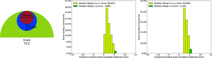
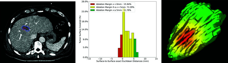
Methods: A QAM computation metric based on volumetric segmentations of tumor and ablation areas and signed Euclidean surface distance maps was developed, including a novel algorithm to address QAM computation in subcapsular tumors. The code for QAM computation was verified in artificial examples of tumor and ablation spheres simulating varying scenarios of ablation margins. The applicability of the QAM metric was investigated in representative cases extracted from a prospective database of colorectal liver metastases (CRLM) treated with stereotactic microwave ablation (SMWA).

Results: Applicability of the proposed QAM metric was confirmed in artificial and clinical example cases. Numerical and visual options of data presentation displaying substrata of QAM distributions were proposed. For subcapsular tumors, the underestimation of tumor coverage by the ablation volume when applying an unadjusted QAM method was confirmed, supporting the benefits of using the proposed algorithm for QAM computation in these cases. The computational code for developed QAM was made publicly available, encouraging the use of a standard and objective metric in reporting ablation completeness and margins.

Conclusion: The proposed volumetric approach for QAM computation including a novel algorithm to address subcapsular liver tumors enables precision and reproducibility in the assessment of ablation margins. The quantitative feedback on ablation completeness opens possibilities for intra-operative decision making and for refined analyses on predictability and consistency of local tumor control after thermal ablation of liver tumors.

Keywords: ablation techniques; computer-assisted therapies; interventional radiology; liver neoplasms; stereotactic techniques.


Conflict of interest statement

SW is co-founder and shareholder of CAScination, the manufacturer of one of the navigation systems applied for stereotactic microwave ablation of colorectal liver metastases in the clinical example cases analyzed in this study. The remaining authors declare that the research was conducted in the absence of any commercial or financial relationships that could be construed as a potential conflict of interest.

Figures

Figure 1
Figure 1
Quantitative ablation margin (QAM) computation pipeline.
Figure 2
Figure 2
Schematic illustration of quantitative ablation margins (QAM) for subcapsular tumors. Left: Subcapsular tumor (red) surrounded by ablation volume (blue). Center: Exclusion volume of 5 mm below liver capsule. Right: Remaining tumor surface used for final QAM computation.
Figure 3
Figure 3
Artificial examples simulating tumor (red) and ablation (blue) volumes. Left: non-subcapsular tumors and ablation volumes. Right: subcapsular tumors with ablation volumes and their relationship to the liver capsule (green).
Figure 4
Figure 4
Visualization of QAM histogram for non-subcapsular artificial case T1.6. Left: artificial example with tumor (red) shifted 5 mm in the x and y directions with respect to the ablation (blue). Right: Histogram of relative distribution of margin distances displayed as 1 mm substrata.
Figure 5
Figure 5
Quantitative ablation margin (QAM) computation for subcapsular artificial case T2.2. with and without applying the algorithm for subcapsular tumors. Left: T2.2 artificial example (tumor: red, ablation: blue, liver: green). Center: Relative distribution of margins with exclusion volume not subtracted in QAM computation. Right: Relative distribution of margins with exclusion volume subtracted in QAM computation.
Figure 6
Figure 6
Volumetric assessment of quantitative ablation margins (QAM) in a patient treated with SMWA for a CRLM in liver segment VI. Left: Computed tomogram image with segmented tumor (red) and ablation (blue) volumes. Center: Relative frequencies of ablation margin substrata with respect to percentage of tumor surface covered. Right: 3D representation of the ablation margin projected onto the ablation surface.
Figure 7
Figure 7
Volumetric assessment of quantitative ablation margins (QAM) in a patient treated with SMWA for a CRLM in liver segment VIII. Left: Computed tomogram image with segmented tumor (red) and ablation (blue) volumes. Center: Relative frequencies of ablation margin substrata with respect to percentage of tumor surface covered. Right: 3D representation of the ablation margin projected onto the ablation surface, showing the area of negative margins in red.
Figure 8
Figure 8
Volumetric assessment of quantitative ablation margins (QAM) of a subcapsular CRLM in liver segment V. Left: Computed tomogram with segmented tumor (red), ablation (blue) volumes and the local liver capsule surrounding the tumor (green). Center: Relative frequencies of ablation margin substrata with respect to percentage of tumor surface covered. Right: 3D representation of the ablation margin projected onto the ablation surface.
Figure 9
Figure 9
Volumetric assessment of quantitative ablation margins (QAM) of a subcapsular CRLM in liver segment VII. Upper left: Computed tomogram with segmented tumor (red), ablation (blue) volumes and the local liver capsule surrounding the tumor (green). Upper right: 3D representation of the ablation margin projected onto the ablation surface. Lower left: Histogram of ablation margin substrata without applying algorithm for subcapsular tumors. Lower right: Relative frequencies of ablation margin substrata with respect to percentage of tumor surface covered when applying algorithm for subcapsular tumors.

Similar articles

Cited by

References

    1. Vasiniotis Kamarinos N, Kaye EA, Sofocleous CT. Image-Guided Thermal Ablation for Colorectal Liver Metastases. Tech Vasc Interv Radiol (2020) 23(2):100672. 10.1016/j.tvir.2020.100672 - DOI - PubMed
    1. Engstrand J, Toporek G, Harbut P, Jonas E, Nilsson H, Freedman J. Stereotactic CT-Guided Percutaneous Microwave Ablation of Liver Tumors With the Use of High-Frequency Jet Ventilation: An Accuracy and Procedural Safety Study. Am J Roentgenol 208(1):193–200. 10.2214/AJR.15.15803 - DOI - PubMed
    1. Heerink WJ, et al. . Robotic versus Freehand Needle Positioning in CT-guided Ablation of Liver Tumors: A Randomized Controlled Trial. Radiology (2017) 290(3):826–32. 10.1148/radiol.2018181698 - DOI - PubMed
    1. Lachenmayer A, Tinguely P, Maurer MH, Frehner L, Knöpfli M, Peterhans M, et al. . Stereotactic image-guided microwave ablation of hepatocellular carcinoma using a computer-assisted navigation system. Liver Int (2019) 39(10):1975–85. 10.1111/liv.14187 - DOI - PubMed
    1. Bale R, Widmann G, Stoffner D, II. Stereotaxy: Breaking the limits of current radiofrequency ablation techniques. Eur J Radiol (2010) 75(1):32–6. 10.1016/j.ejrad.2010.04.013 - DOI - PubMed

LinkOut - more resources