Clinical Trial
. 2021 Mar 10:2021:6644897.
doi: 10.1155/2021/6644897. eCollection 2021.

Association of Matrix Metalloproteinase-2 mRNA Expression with Subtypes of Pediatric Cholesteatoma

Affiliations
Clinical Trial

Association of Matrix Metalloproteinase-2 mRNA Expression with Subtypes of Pediatric Cholesteatoma

Taichi Kan et al. Biomed Res Int. .

Abstract

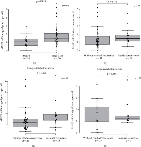
Objective: Cholesteatoma is a clinically heterogeneous disease, with some patients showing spontaneous regression, while others experiencing an aggressive, lethal disease. Cholesteatoma in children can be divided into two types: congenital and acquired. Identifying good prognostic markers is needed to help select patients who will require immediate surgical intervention. Matrix metalloproteinase-2 (MMP2) was previously reported to play an important role in cholesteatoma progression, by promoting bone destruction and keratinocyte infiltration. Herein, we analyzed MMP2 mRNA expression level in cholesteatoma using RNA-in situ hybridization in formalin-fixed, paraffin-embedded (FFPE) tissue samples.

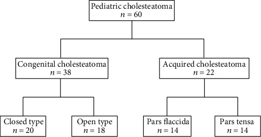
Methods: Sixty patients with cholesteatoma under 15 years old, who underwent their primary surgery at Aichi Medical University's Otolaryngology Department, were analyzed for MMP2 expression level, using RNA-in situ hybridization.

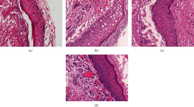
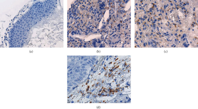
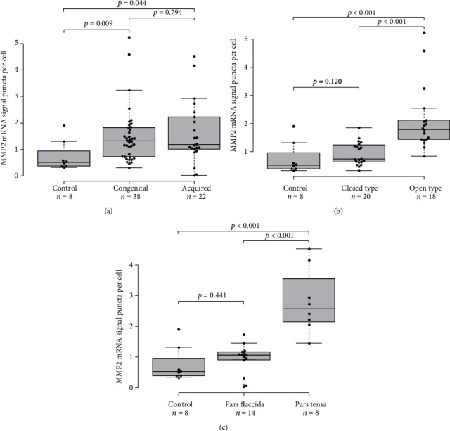
Results: There were no significant differences in MMP2 mRNA expression level between congenital cholesteatoma and acquired cholesteatomas. In congenital cholesteatoma, higher MMP2 signals were observed in the open type than in the closed type (p < 0.001). In acquired cholesteatoma, higher MMP2 signals were observed in the pars tensa than in the pars flaccida (p < 0.001). MMP2 mRNA expression level was almost exclusively found in the fibroblasts or in the inflammatory cells in the stroma, but not in the epithelium.

Conclusion: Our study reveals that MMP2 mRNA expression level is strongly associated with the subtypes of cholesteatoma. The findings suggest that the level of expression of MMP2 mRNA may be related to the pathogenesis and aggressive features of cholesteatoma.


Conflict of interest statement

The authors have no significant relationships with or financial interests in any commercial companies pertaining to this article.

Figures

Figure 1
Figure 1
Flowchart of patient selection.
Figure 2
Figure 2
Representative histology of pediatric cholesteatoma. (a) Congenital cholesteatoma, the closed type showing a cystic structure that is lined by squamous epithelium and thin fibrous stroma with a scarce amount of inflammatory cells. (b) Congenital cholesteatoma, the open type showing a cystic structure that is lined by squamous epithelium and fibrous connective tissue with inflammation. (c) Acquired cholesteatoma, the pars flaccida type showing a cystic structure, squamous epithelium, and stroma with fibrosis. (d) The pars tensa type showing a cystic structure, squamous epithelium, and thick stroma with plasma cell-predominant inflammatory cells.
Figure 3
Figure 3
RNA-in situ hybridization assay in pediatric cholesteatoma, probing for MMP2. (a) A minute number of MMP2-positive signals were observed in congenital cholesteatoma (closed type). (b–d) MMP2-positive signals were observed in inflammatory cells in the stroma of congenital cholesteatoma (open type), pars flaccida type, and pars tensa type of acquired cholesteatoma. The MMP2 signal is shown as brown puncta.
Figure 4
Figure 4
Box-and-whisker plot showing MMP2 mRNA expression levels in cholesteatomas. The MMP2 mRNA expression level was quantified as the average number of DAB signal puncta per cell using Vectra 3 (PerkinElmer Co, Waltham, MA). The signals and cells within the shooting range were automatically counted. The average numbers between subtypes of cholesteatoma and normal skin were compared using the Mann–Whitney U test, and p values are presented as a box-and-whisker plot. The means with standard error of the means (SEM) are shown for each group. (a) A comparison of MMP2 mRNA expression levels in congenital cholesteatoma, acquired cholesteatoma, and normal skin from the accessory ear (shown with control). There was no significant difference in the mRNA expression level of MMP2 between congenital cholesteatoma and acquired cholesteatoma. Congenital cholesteatoma and acquired cholesteatoma had significantly higher MMP2 mRNA expression levels than the normal skin (p = 0.009 and p = 0.044, respectively). (b) A comparison of MMP2 mRNA expression levels between closed and open types of congenital cholesteatoma. The open type had higher MMP2 mRNA expression levels than the closed type (p < 0.001) and normal skin (p < 0.001). No significant difference was observed between the closed type and normal skin. (c) A comparison of MMP2 mRNA expression levels between the pars flaccida and pars tensa types of acquired cholesteatoma. The pars tensa type had significantly higher MMP2 mRNA expression levels than the pars flaccida type (p < 0.001) and normal skin (p < 0.001). No significant difference was observed between the pars flaccida type and normal skin.
Figure 5
Figure 5
Box-and-whisker plot showing the association of MMP2 mRNA expression levels with clinical features. (a) A comparison of MMP2 mRNA expression levels between cholesteatomas from stage I and stage II or III. Significantly higher expression levels were observed in stage II or stage III group (p = 0.019). (b) A comparison of MMP2 mRNA expression levels between cholesteatomas in this study with and without residual or recurrent lesions (p = 0.174). (c) A comparison of MMP2 mRNA expression levels between congenital cholesteatoma with and without residual or recurrent lesions (p = 0.125). (d) A comparison of MMP2 mRNA expression levels between congenital cholesteatoma with and without residual or recurrent lesions. No apparent trend was observed between the two groups (p = 0.695).

Similar articles

Cited by

References

    1. Maniu A., Harabagiu O., Perde Schrepler M., Catana A., Fanuta B., Mogoanta C. A. Molecular biology of cholesteatoma. Romanian Journal of Morphology and Embryology. 2014;55(1):7–13. - PubMed
    1. Aimi K. Role of the tympanic ring in the pathogenesis of congenital cholesteatoma. Laryngoscope. 1983;93(9):1140–1146. doi: 10.1288/00005537-198309000-00005. - DOI - PubMed
    1. Michaels L. An epidermoid formation in the developing middle ear: possible source of cholesteatoma. The Journal of Otolaryngology. 1986;15(3):169–174. - PubMed
    1. Michaels L. Origin of congenital cholesteatoma from a normally occurring epidermoid rest in the developing middle ear. International Journal of Pediatric Otorhinolaryngology. 1988;15(1):51–65. doi: 10.1016/0165-5876(88)90050-X. - DOI - PubMed
    1. Kodama K., Hara M., Hasegawa M., et al. Two cases of spontaneous regression of congenital cholesteatomas. International Journal of Pediatric Otorhinolaryngology. 2012;76(1):142–144. doi: 10.1016/j.ijporl.2011.07.008. - DOI - PubMed

Publication types

Supplementary concepts

LinkOut - more resources