

Construction of heparin-based hydrogel incorporated with Cu5.4O ultrasmall nanozymes for wound healing and inflammation inhibition
- PMID: 33778192
- PMCID: PMC7960791
- DOI: 10.1016/j.bioactmat.2021.02.006
Construction of heparin-based hydrogel incorporated with Cu5.4O ultrasmall nanozymes for wound healing and inflammation inhibition
Erratum in
-
Erratum regarding missing ethics approval and consent to participate statements in previously published articles.Bioact Mater. 2024 Jun 14;40:275-279. doi: 10.1016/j.bioactmat.2024.06.006. eCollection 2024 Oct. Bioact Mater. 2024. PMID: 38973994 Free PMC article.
Abstract
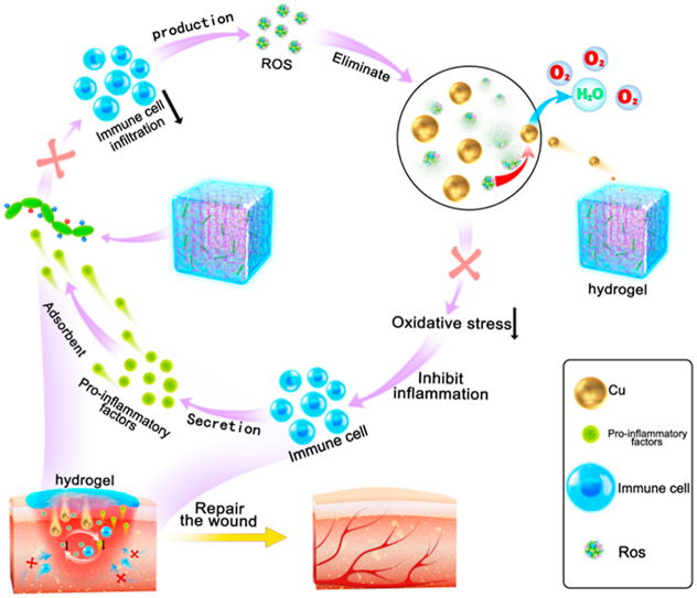
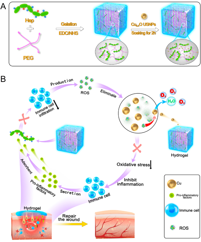
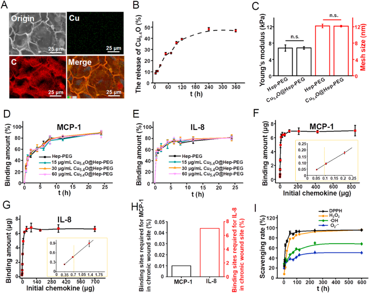
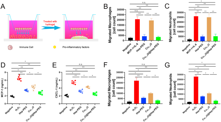
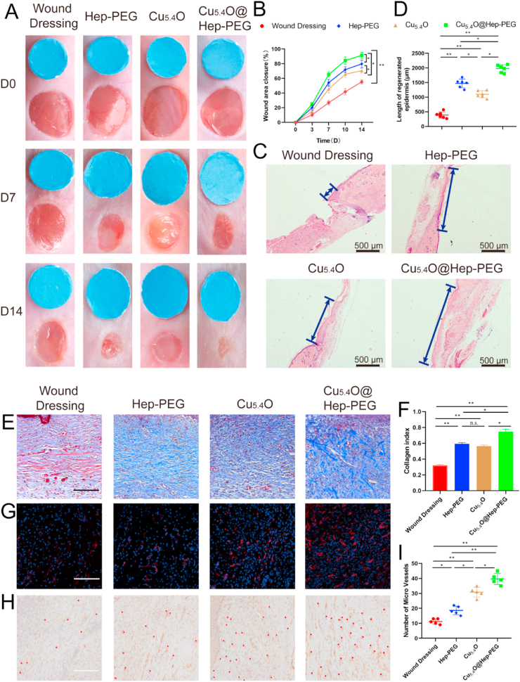
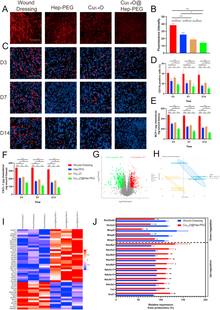
Excessive production of inflammatory chemokines and reactive oxygen species (ROS) can cause a feedback cycle of inflammation response that has a negative effect on cutaneous wound healing. The use of wound-dressing materials that simultaneously absorb chemokines and scavenge ROS constitutes a novel 'weeding and uprooting' treatment strategy for inflammatory conditions. In the present study, a composite hydrogel comprising an amine-functionalized star-shaped polyethylene glycol (starPEG) and heparin for chemokine sequestration as well as Cu5.4O ultrasmall nanozymes for ROS scavenging (Cu5.4O@Hep-PEG) was developed. The material effectively adsorbs the inflammatory chemokines monocyte chemoattractant protein-1 and interleukin-8, decreasing the migratory activity of macrophages and neutrophils. Furthermore, it scavenges the ROS in wound fluids to mitigate oxidative stress, and the sustained release of Cu5.4O promotes angiogenesis. In acute wounds and impaired-healing wounds (diabetic wounds), Cu5.4O@Hep-PEG hydrogels outperform the standard-of-care product Promogram® in terms of inflammation reduction, increased epidermis regeneration, vascularization, and wound closure.
Keywords: Hydrogels; Inflammatory chemokines; Nanozymes; Reactive oxygen species; Wound healing.
© 2021 The Authors.
Conflict of interest statement
The authors have no competing financial interests or personal relationships that could influence the work published in this paper.
Figures
Similar articles
-
Ultrasmall Antioxidant Copper Nanozyme to Enhance Stem Cell Microenvironment for Promoting Diabetic Wound Healing.Int J Nanomedicine. 2024 Dec 19;19:13563-13578. doi: 10.2147/IJN.S487647. eCollection 2024. Int J Nanomedicine. 2024. PMID: 39720217 Free PMC article.
-
Glycosaminoglycan-based hydrogels capture inflammatory chemokines and rescue defective wound healing in mice.Sci Transl Med. 2017 Apr 19;9(386):eaai9044. doi: 10.1126/scitranslmed.aai9044. Sci Transl Med. 2017. PMID: 28424334
-
Construction of programmed time-released multifunctional hydrogel with antibacterial and anti-inflammatory properties for impaired wound healing.J Nanobiotechnology. 2024 Mar 23;22(1):126. doi: 10.1186/s12951-024-02390-y. J Nanobiotechnology. 2024. PMID: 38519957 Free PMC article.
-
Integrated cascade antioxidant nanozymes-Cu5.4O@CNDs combat acute liver injury by regulating retinol metabolism.Theranostics. 2025 Apr 21;15(12):5592-5615. doi: 10.7150/thno.106811. eCollection 2025. Theranostics. 2025. PMID: 40365282 Free PMC article.
-
Multifunctional and theranostic hydrogels for wound healing acceleration: An emphasis on diabetic-related chronic wounds.Environ Res. 2023 Dec 1;238(Pt 1):117087. doi: 10.1016/j.envres.2023.117087. Epub 2023 Sep 15. Environ Res. 2023. PMID: 37716390 Review.
Cited by
-
Copper incorporated biomaterial-based technologies for multifunctional wound repair.Theranostics. 2024 Jan 1;14(2):547-570. doi: 10.7150/thno.87193. eCollection 2024. Theranostics. 2024. PMID: 38169658 Free PMC article. Review.
-
Functional drug-delivery hydrogels for oral and maxillofacial wound healing.Front Bioeng Biotechnol. 2023 Aug 3;11:1241660. doi: 10.3389/fbioe.2023.1241660. eCollection 2023. Front Bioeng Biotechnol. 2023. PMID: 37600316 Free PMC article. Review.
-
A Review of Immunomodulatory Reprogramming by Probiotics in Combating Chronic and Acute Diabetic Foot Ulcers (DFUs).Pharmaceutics. 2022 Nov 10;14(11):2436. doi: 10.3390/pharmaceutics14112436. Pharmaceutics. 2022. PMID: 36365254 Free PMC article. Review.
-
Insights into the Role of Natural Polysaccharide-Based Hydrogel Wound Dressings in Biomedical Applications.Gels. 2022 Oct 12;8(10):646. doi: 10.3390/gels8100646. Gels. 2022. PMID: 36286147 Free PMC article. Review.
-
Efficient angiogenesis-based wound healing through hydrogel dressing with extracellular vesicles release.Mater Today Bio. 2022 Sep 24;16:100427. doi: 10.1016/j.mtbio.2022.100427. eCollection 2022 Dec. Mater Today Bio. 2022. PMID: 36193344 Free PMC article.
References
-
- Singer A.J., Clark R.A. Cutaneous wound healing. N. Engl. J. Med. 1999;1:738–746. - PubMed
-
- Armstrong D.G., Boulton A.J.M., Bus S.A. Diabetic foot ulcers and their recurrence. N. Engl. J. Med. 2017;376(24):2367–2375. - PubMed
-
- Kurita M., Araoka T., Hishida T., O'Keefe D.D., Takahashi Y., Sakamoto A., Sakurai M., Suzuki K., Wu J., Yamamoto M., Hernandez-Benitez R., Ocampo A., Reddy P., Shokhirev M.N., Magistretti P., Delicado E.N., Eto H., Harii K., Belmonte J.C.I. In vivo reprogramming of wound-resident cells generates skin epithelial tissue. Nature. 2018;561(7722):243–247. - PMC - PubMed
-
- Shiekh P.A., Singh A., Kumar A. Exosome laden oxygen releasing antioxidant and antibacterial cryogel wound dressing OxOBand alleviate diabetic and infectious wound healing. Biomaterials. 2020;249:120020. - PubMed
-
- Tang Q., Lim T., Wei X.J., Wang Q.Y., Xu J.C., Shen L.Y., Zhu Z.Z., Zhang C.Q. A free-standing multilayer film as a novel delivery carrier of platelet lysates for potential wound-dressing applications. Biomaterials. 2020;255:120138. - PubMed
LinkOut - more resources
Full Text Sources
Other Literature Sources
Research Materials
