. 2021 Mar 13;6(10):3288-3299.
doi: 10.1016/j.bioactmat.2021.02.035. eCollection 2021 Oct.

l-cysteine-modified chiral gold nanoparticles promote periodontal tissue regeneration

Affiliations

l-cysteine-modified chiral gold nanoparticles promote periodontal tissue regeneration

Shuang Zhang et al. Bioact Mater. .

Abstract

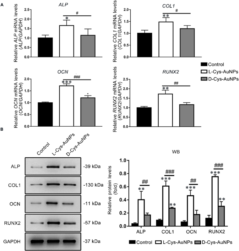
Gold nanoparticles (AuNPs) with surface-anchored molecules present tremendous potential in tissue regeneration. However, little is known about chiral-modified AuNPs. In this study, we successfully prepared L/D-cysteine-anchored AuNPs (L/D-Cys-AuNPs) and studied the effects of chiral-modified AuNPs on osteogenic differentiation and autophagy of human periodontal ligament cells (hPDLCs) and periodontal tissue regeneration. In vitro, more L-Cys-AuNPs than D-Cys-AuNPs tend to internalize in hPDLCs. L-Cys-AuNPs also significantly increased the expression of alkaline phosphatase, collagen type 1, osteocalcin, runt-related transcription factor 2, and microtubule-associated protein light chain 3 II and decreased the expression of sequestosome 1 in hPDLCs compared to the expression levels in the hPDLCs treated by D-Cys-AuNPs. In vivo tests in a rat periodontal-defect model showed that L-Cys-AuNPs had the greatest effect on periodontal-tissue regeneration. The activation of autophagy in L-Cys-AuNP-treated hPDLCs may be responsible for the cell differentiation and tissue regeneration. Therefore, compared to D-Cys-AuNPs, L-Cys-AuNPs show a better performance in cellular internalization, regulation of autophagy, cell osteogenic differentiation, and periodontal tissue regeneration. This demonstrates the immense potential of L-Cys-AuNPs for periodontal regeneration and provides a new insight into chirally modified bioactive nanomaterials.

Keywords: Autophagy; Chirality; Gold nanoparticles; Periodontal; Tissue regeneration.


Conflict of interest statement

None.

Figures

Image 1
Graphical abstract
Fig. 1
Fig. 1
Characterization of bare AuNPs, L-Cys-AuNPs, and D-Cys-AuNPs. (A) TEM images. Inset: corresponding photographs. (B) DLS images. (C) CD spectra. (D) UV–vis absorption spectra. (E) Zeta potential of the samples in water and 10% FBS. ***P < 0.001 compared with the control group, ###P < 0.001.
Fig. 2
Fig. 2
Biocompatibility, uptake, cellular localization, ALP activity, and mineral deposition of L/D-Cys-AuNPs. hPDLCs were cultured with AuNPs. (A) Light microscopy images of hPDLCs before and after treatment with different types of AuNPs for 3 days. (B) Effects of AuNPs on cell viability and proliferation of hPDLCs on days 1, 3, 5, and 7. (C) TEM images of hPDLCs incubated with AuNPs on day 3. The arrows indicate the internalized AuNPs. (D) Concentrations of Au on day 3. ND means not detected. (E) ALP activity levels on days 3, 5, and 7. (F) ALP staining on day 7. (G) ARS and von Kossa staining and quantification of mineral deposition on day 21. *P < 0.05, **P < 0.01, ***P < 0.001 compared with the control group, #P < 0.05, ##P < 0.01, ###P < 0.001.
Fig. 3
Fig. 3
Effects of L/D-Cys-AuNPs on the osteogenic genes and proteins of hPDLCs cultured with L/D-Cys-AuNPs. (A) ALP, COL1, OCN and RUNX2 mRNA levels of L/D-Cys-AuNP-treated hPDLCs on day 5. (B) ALP, COL1, OCN and RUNX2 protein levels of L/D-Cys-AuNP-treated hPDLCs on day 7. *P < 0.05, **P < 0.01, ***P < 0.001 compared with the control group, #P < 0.05, ##P < 0.01, ###P < 0.001.
Fig. 4
Fig. 4
Effects of L/D-Cys-AuNPs on autophagy genes and proteins of hPDLCs cultured with L/D-Cys-AuNPs. (A) BECN1 and LC3 mRNA levels of L/D-Cys-AuNP-treated hPDLCs on days 3, 5, and 7. (B) LC3-II and SQSTM1 protein levels of L/D-Cys-AuNP-treated hPDLCs on day 7. hPDLCs were then cultured with the autophagy inhibitor 3-MA and L/D-Cys-AuNPs. (C) ALP staining on day 7 and ARS staining on day 21. (D) COL1, RUNX2, and ALP protein levels of 3-MA and L/D-Cys-AuNP-treated hPDLCs on day 7. *P < 0.05, **P < 0.01, ***P < 0.001 compared with the control group, #P < 0.05, ##P < 0.01, ###P < 0.001.
Fig. 5
Fig. 5
L-Cys-AuNPs showed the best in-vivo periodontal regeneration potential in the rat periodontal-fenestration-defect model. hPDLCs were seeded onto a resorbable collagen membrane and cultured with L/D-Cys-AuNPs. The treated cell-membrane compounds were placed into the rat periodontal-fenestration-defect models and embedded for 21 days. (A) μCT images of the mandibular first molars (B means buccal and L means lingual). (B) Bone-related parameters (BMD, TV, BV, BV/TV, Tb.Th, and Tb.N) reported by μCT. (C) Quantification of NAB and NPDL. (D) The representative H&E, Masson, VG, and Goldner's staining images (D means dentin, NAB means newly-formed alveolar bones, and NPDL means newly-formed periodontal ligaments). *P < 0.05, **P < 0.01, ***P < 0.001 compared with the control group, #P < 0.05.
Fig. 6
Fig. 6
L/D-Cys-AuNP-modulated autophagy in the rat periodontal-fenestration-defect model. hPDLCs were seeded onto a resorbable collagen membrane and cultured with L/D-Cys-AuNPs. The treated cell-membrane compounds were placed into the rat periodontal-fenestration-defect models and cultured for 21 days. (A) Expression of LC3 and SQSTM1 protein levels in the periodontal tissue of the mandibular first molars (D means dentin). (B) Quantification of LC3 and SQSTM1. *P < 0.05, **P < 0.01 compared with the control group.

References

    1. Pihlstrom B.L., Michalowicz B.S., Johnson N.W. Periodontal diseases. Lancet. 2005;366(9499):1809–1820. - PubMed
    1. Offenbacher S., Jiao Y., Kim S.J., Marchesan J., Moss K.L., Jing L., Divaris K., Bencharit S., Agler C.S., Morelli T., Zhang S., Sun L., Seaman W.T., Cowley D., Barros S.P., Beck J.D., Munz M., Schaefer A.S., North K.E. GWAS for Interleukin-1beta levels in gingival crevicular fluid identifies IL37 variants in periodontal inflammation. Nat. Commun. 2018;9(1):3686. - PMC - PubMed
    1. Seo B.-M., Miura M., Gronthos S., Mark Bartold P., Batouli S., Brahim J., Young M., Gehron Robey P., Wang C.Y., Shi S. Investigation of multipotent postnatal stem cells from human periodontal ligament. Lancet. 2004;364(9429):149–155. - PubMed
    1. Zhou J., Zhang Y., Li L., Fu H., Yang W., Yan F. Human beta-defensin 3-combined gold nanoparticles for enhancement of osteogenic differentiation of human periodontal ligament cells in inflammatory microenvironments. Int. J. Nanomed. 2018;13:555–567. - PMC - PubMed
    1. Zhang Y., Kong N., Zhang Y., Yang W., Yan F. Size-dependent effects of gold nanoparticles on osteogenic differentiation of human periodontal ligament progenitor cells. Theranostics. 2017;7(5):1214–1224. - PMC - PubMed

LinkOut - more resources