. 2021 Jan 3;5(3):e10454.
doi: 10.1002/jbm4.10454. eCollection 2021 Mar.

Abnormal Bone Collagen Cross-Linking in Osteogenesis Imperfecta/Bruck Syndrome Caused by Compound Heterozygous PLOD2 Mutations

Affiliations

Abnormal Bone Collagen Cross-Linking in Osteogenesis Imperfecta/Bruck Syndrome Caused by Compound Heterozygous PLOD2 Mutations

Charlotte Gistelinck et al. JBMR Plus. .

Abstract

Bruck syndrome (BS) is a congenital disorder characterized by joint flexion contractures, skeletal dysplasia, and increased bone fragility, which overlaps clinically with osteogenesis imperfecta (OI). On a genetic level, BS is caused by biallelic mutations in either FKBP10 or PLOD2. PLOD2 encodes the lysyl hydroxylase 2 (LH2) enzyme, which is responsible for the hydroxylation of cross-linking lysine residues in fibrillar collagen telopeptide domains. This modification enables collagen to form chemically stable (permanent) intermolecular cross-links in the extracellular matrix. Normal bone collagen develops a unique mix of such stable and labile lysyl-oxidase-mediated cross-links, which contribute to bone strength, resistance to microdamage, and crack propagation, as well as the ordered deposition of mineral nanocrystals within the fibrillar collagen matrix. Bone from patients with BS caused by biallelic FKBP10 mutations has been shown to have abnormal collagen cross-linking; however, to date, no direct studies of human bone from BS caused by PLOD2 mutations have been reported. Here the results from a study of a 4-year-old boy with BS caused by compound heterozygous mutations in PLOD2 are discussed. Diminished hydroxylation of type I collagen telopeptide lysines but normal hydroxylation at triple-helical sites was found. Consequently, stable trivalent cross-links were essentially absent. Instead, allysine aldol dimeric cross-links dominated as in normal skin collagen. Furthermore, in contrast to the patient's bone collagen, telopeptide lysines in cartilage type II collagen cross-linked peptides from the patient's urine were normally hydroxylated. These findings shed light on the complex mechanisms that control the unique posttranslational chemistry and cross-linking of bone collagen, and how, when defective, they can cause brittle bones and related connective tissue problems. © 2020 The Authors. JBMR Plus published by Wiley Periodicals LLC. on behalf of American Society for Bone and Mineral Research.

Keywords: BRUCK SYNDROME; COLLAGEN; LYSYL HYDROXYLASE 2; OSTEOGENESIS IMPERFECTA; PLOD2.


Figures

Fig 1
Fig 1
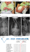
Clinical and molecular studies. (A) Photographs taken at age 4 months of the proband showing contractures of the joints. (B) Radiographs of proband at birth and 10 years, and of the affected sibling at the age of 8 years. Radiographs of proband show multiple rib fractures and platyspondyly at birth and the fixation of cervical and upper thoracic spine at the age of 10 years. The affected sibling shows scoliosis, partially corrected by surgical rodding at age 8 years. (C) Schematic representation of the genomic structure of the H. sapiens PLOD2 gene. Boxes and lines represent exons and introns, respectively. Exon 13a (thick box border) is subject to differential splicing. The catalytic domain is encoded by the last four exons of PLOD2 (indicated by the green dashed line). One mutation in the proband results in a stop‐codon in exon 12 of PLOD2 (p.Arg440Ter), whereas the other allele carries a mutation that substitutes a highly conserved Pro residue in exon 17 of PLOD2 (p.Pro653Arg). The location of this mutation, which is a part of the catalytic domain, represents a mutation hotspot( 37 , 38 , 41 ); the substitution was predicted to be deleterious in silico (polyPhen). HP = hydroxy lysyl pyridinoline; LP = lysyl pyridinoline.
Fig 2
Fig 2
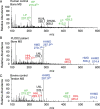
Mass‐spectral analysis of a type II collagen cross‐linked peptide from urine. Patient's urine was fractionated and analyzed by liquid chromatography‐mass spectrometry. Shown is a peptide identified in this sample, which comes from a site of α1(II) C‐telopeptide pyridinoline cross‐linking (M = 1576), reported earlier to be present in control children's urine.( 20 ) The parent ion envelope of this HPyr‐containing structure is shown, whereas the posttranslational variant LPyr‐containing cross‐link (486.63+) is only present in very low amounts. This result shows that in contrast to bone type I collagen, the patient's cartilage type II collagen had apparently normal telopeptide lysine hydroxylation, and the low level of the LPyr form of the peptide indicates almost complete hydroxylation of the triple‐helical domain lysines in tissue from which the peptides originated.
Fig 3
Fig 3
SDS‐PAGE analysis of bone collagen. (A) Collagen extracted from fetal control, adult control, and patient's bone was denatured and run on SDS‐PAGE. The pattern of collagen chains extracted from patient's bone is more similar to that of normal skin than bone collagen, with more prominent β‐dimers (red arrowhead) and γ‐trimers (black arrowhead). This is consistent with cross‐linking by lysine aldehydes not hydroxylysine aldehydes. (B) Nondenaturing extraction in dilute acetic acid solubilizes little collagen from normal fetal or adult demineralized bone, whereas the patient's bone collagen is highly extractable, consistent with a low level of acid‐stable intermolecular cross‐links.
Fig 4
Fig 4
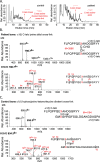
Mass‐spectral analysis of the effect of the PLOD2 defect on collagen I cross‐linking. Liquid chromatography‐mass spectrometry (LC‐MS) fractionation of collagenase‐digested bone collagen peptides from normal control bone and patient's bone. (A) The chromatogram of the patient's bone reveals an increase in the presence of a linear fragment corresponding to the C‐telopeptide of type I collagen (C‐telo linear, confirmed by MS), and an allysine aldol cross‐linked C‐telopeptide dimer (C‐telo lysine divalent cross‐link), which is a minor component of normal bone. (B) MS confirmed the peptide dimer structure matched the mass of an allysine aldol cross‐link between two α1(I) C‐telopeptides (structure shown, M = 3986 Da). The parent ion envelope of the latter (665.96+, 798.75+, 998.04+, etc) is evident in the MS spectrum, and the MS/MS fragmentation pattern shown below confirms the two peptide sequences present. (C) As a reference, LC‐MS of a digest of normal bone collagen shows in contrast a prominent divalent cross‐linking structure formed between the α1(I) C‐telopeptide of one molecule and the α1(I) helical domain of another molecule, in which the cross‐link has the mass of the galactosyl form of hydroxylysino‐ketonorleucine. The charge envelope of the parent ion (M = 4164), and the MS/MS fragmentation pattern shown below, confirms the structure, which is the precursor for trivalent pyridinoline and pyrrole cross‐links in bone collagen.
Fig 5
Fig 5
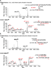
Mass‐spectral identification of N‐telopeptide aldol cross‐linked structures in the patient's bone. (A) From fraction 36 of the chromatogram of the collagenase‐digested patient's bone collagen shown in Fig. 4A (right panel), we also recovered on liquid chromatography‐mass spectrometry (LC‐MS) analysis a prominent aldol‐cross‐linked α1(I)N to α2(I) N‐telopeptide dimer. Upper panel: The MS spectrum shows the parent ion envelope for M = 2855, which is the mass of the structure shown. Lower panel: The parent ion MSMS fragmentation pattern confirms the structure. (B) In contrast with normal bone, the equal mix of hydroxylysine and lysine aldehydes produces divalent keto‐imine intermolecular cross‐links that can mature to trivalent pyridinolines and pyrroles. The peptide shown was prominent on LC‐MS analysis of bacterial collagenase‐digested control bone matrix (Fig. 4A , left panel). It originates from a divalent intermolecular hydroxylysino‐ketonorleucine cross‐link between an α2(I) N‐telopeptide and α2(I) K933. Upper panel: The MS spectrum shows the parent ion envelope for M = 2578, the mass of the structure containing this cross‐link. Lower panel: The parent ion MSMS fragmentation pattern confirms the peptide structure.
Fig 6
Fig 6
Liquid chromatography‐mass spectrometry (LC‐MS) analysis of collagen cross‐link derivatives in acid hydrolysates of borohydride‐reduced PLOD2‐mutant bone compared with control bone and tendon. (A) From control bone, the pyridinoline (Pyr) cross‐links (green) and their divalent precursors, DHLNL and HLNL, predominate. From PLOD2‐mutant bone, Pyrs are absent and histidinohydroxymerodesmosine (HHMD; and histidinomerodesmosine [HMD]) predominate with small amounts of histidinohydroxylysinonorleucine (HHL). (B) HHMD and HHL are more typical of skin collagen and are the artifactual products of allysine aldol telopeptide dimers on acid hydrolysis (HHL from C‐telopeptide aldols) and borohydride reduction (HHMD from N‐telopeptide aldols).( 29 ) The lysyl homolog of HHMD (HMD) is prominent in PLOD2‐mutant bone collagen because the helical‐domain cross‐linking lysines at α1(I)K930 and α2(I)K933 are only partially hydroxylated in normal and PLOD2 bone. (C) For comparison, results are shown from bovine tendon collagen, a tissue that also expresses a mix of telopeptide lysine and hydroxylysine aldehyde cross‐links.
Fig 7
Fig 7
Hydroxylation status of telopeptide cross‐linking lysine residues in type I collagen. Linear (uncross‐linked) peptides from the N‐ and C‐telopeptide domains of collagen α‐chains were prepared by in‐gel trypsin digestion (N‐telopeptide) and bacterial collagenase digestion (C‐telopeptide) of decalcified bone collagen and identified by liquid chromatography‐mass spectrometry (LC‐MS). Right panels: Informative peptides recovered from control bone (upper) and the patient's (lower) bone showed 47% and 0% hydroxylation, respectively. Left panels: The patient's bone yielded only an unhydroxylated α1(I) N‐telopeptide; upper: MS of parent ion; lower: MSMS fragment identification. See Table 2 for yields. No informative α2(I)N‐telopeptide was recovered.
Fig 8
Fig 8
Hydroxylation status of triple‐helical domain cross‐linking lysine residues in type I collagen. Linear (uncross‐linked) peptides from the four molecular sites of cross‐linking in the helical domain of collagen type I were also prepared for liquid chromatography‐mass spectrometry (LC‐MS) analysis by bacterial collagenase digestion of decalcified bone. Results are shown on linear sequences containing residues K87 and K930 from the α1(I)‐chain. The K87 site was 95% hydroxylated from both the normal control's and the patient's bone, of which most was glycosylated primarily as K * gal for both. The K930 site was 40% hydroxylated in control bone and 34% in the patient's bone with no glycosylation (ie, both in the normal bone range). This peptide has two hydroxylatable lysines so the parent ion +16 ladder reflects their status shown as KK, KK*, and K*K*. The hydroxylation percentages in the figure refer specifically to residue K930, based on validation by MSMS fragmentation of the individual parent ions (not shown). See Table 2 for a summary of hydroxylation levels at all helical cross‐linking Lys sites.
Fig 9
Fig 9
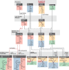
Schematic of the mixed function cross‐linking from lysine and hydroxylysine aldehydes in normal bone collagen to illustrate the effect of lysine aldehydes alone in PLOD2 bone. In normal human bone type I collagen, approximately 50% of the telopeptide lysines (in red) are converted by PLOD2/LH2 to telopeptide hydroxylysines (in yellow). Both are substrates for lysyl oxidase (LOX)–producing reactive aldehydes (allysines) extracellularly, which react with the specific lysine or hydroxylysine side chains (in green and blue respectively) in triple‐helical domains of adjacent molecules forming keto‐imine and aldimine divalent cross‐links. A fraction of the latter goes on to interact, forming permanent trivalent cross‐links (pyrroles and pyridinolines). From 50% telopeptide hydroxylysine (in yellow) at synthesis, approximately equal amounts of pyrroles and pyridinolines would result.( 22 ) Red dashed lines summarize the effect of PLOD2/LH2 deficiency, with fewer or no trivalent and stable divalent cross‐links forming and allysine aldol dimers the main products. The latter on tissue borohydride reduction produce the complex structures HHMD (histidinohydroxymerodesmosine) and HMD (histidinomerodesmosine; Fig. 6), and on acid hydrolysis, the artifact HHL (histidinohydroxylysinonorleucine; see Eyre and colleagues( 29 ) for details). We speculate that the material strength and toughness of the collagen as a mineralized bone composite are impaired by the labile nature of the dermis‐like cross‐linking.

References

    1. Eyre DR, Weis MA. Bone collagen: new clues to its mineralization mechanism from recessive osteogenesis imperfecta. Calcif Tissue Int. 2013;93(4):338–47. - PMC - PubMed
    1. Nyman JS, Reyes M, Wang X. Effect of ultrastructural changes on the toughness of bone. Micron. 2005;36(7–8):566–82. - PubMed
    1. Nyman JS, Zhai H, Bigio EH, et al. Age‐related factors affecting the postyield energy dissipation of human cortical bone. J Orthop Res. 2007;25(5):646–55. - PMC - PubMed
    1. Wu JJ, Weis MA, Kim LS, et al. Differences in chain usage and cross‐linking specificities of cartilage type V/XI collagen isoforms with age and tissue. J Biol Chem. 2009;284(9):5539–45. - PMC - PubMed
    1. Mercer DK, Nicol PF, Kimbembe C, Robins SP. Identification, expression, and tissue distribution of the three rat lysyl hydroxylase isoforms. Biochem Biophys Res Commun. 2003;307(4):803–9. - PubMed

LinkOut - more resources