

Assessing Risk of Breast Cancer: A Review of Risk Prediction Models
- PMID: 33778488
- PMCID: PMC7980704
- DOI: 10.1093/jbi/wbab001
Assessing Risk of Breast Cancer: A Review of Risk Prediction Models
Abstract
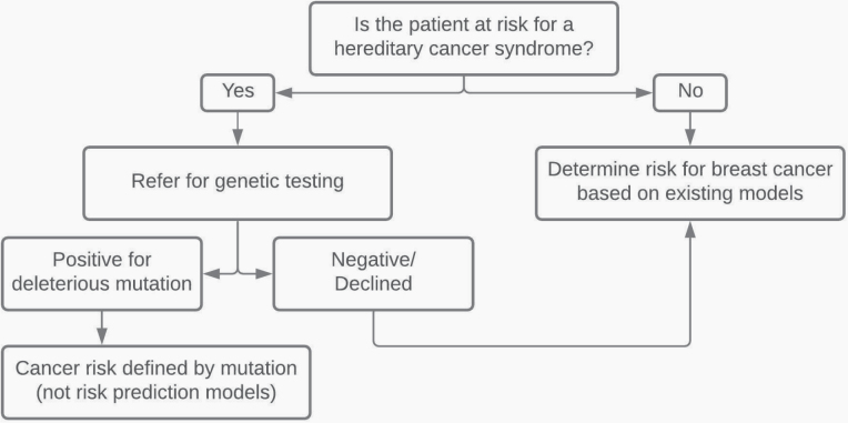
Accurate and individualized breast cancer risk assessment can be used to guide personalized screening and prevention recommendations. Existing risk prediction models use genetic and nongenetic risk factors to provide an estimate of a woman's breast cancer risk and/or the likelihood that she has a BRCA1 or BRCA2 mutation. Each model is best suited for specific clinical scenarios and may have limited applicability in certain types of patients. For example, the Breast Cancer Risk Assessment Tool, which identifies women who would benefit from chemoprevention, is readily accessible and user-friendly but cannot be used in women under 35 years of age or those with prior breast cancer or lobular carcinoma in situ. Emerging research on deep learning-based artificial intelligence (AI) models suggests that mammographic images contain risk indicators that could be used to strengthen existing risk prediction models. This article reviews breast cancer risk factors, describes the appropriate use, strengths, and limitations of each risk prediction model, and discusses the emerging role of AI for risk assessment.
Keywords: breast cancer; mammography; risk assessment; screening.
© Society of Breast Imaging 2021. All rights reserved. For permissions, please e-mail: journals.permissions@oup.com.
Figures
References
-
- Saslow D, Boetes C, Burke W, et al. ; American Cancer Society Breast Cancer Advisory Group . American Cancer Society guidelines for breast screening with MRI as an adjunct to mammography. CA Cancer J Clin 2007;57(2):75–89. - PubMed
-
- Monticciolo DL, Newell MS, Moy L, Niell B, Monsees B, Sickles EA. Breast cancer screening in women at higher-than-average risk: recommendations from the ACR. J Am Coll Radiol 2018;15(3):408–414. - PubMed
-
- Bakker MF, de Lange SV, Pijnappel RM, et al. ; DENSE Trial Study Group . Supplemental MRI screening for women with extremely dense breast tissue. N Engl J Med 2019;381(22):2091–2102. - PubMed
-
- Wang L, Strigel RM. Supplemental screening for patients at intermediate and high risk for breast cancer. Radiol Clin North Am 2021;59(1):67–83. - PubMed
Publication types
Grants and funding
LinkOut - more resources
Full Text Sources
Other Literature Sources
Miscellaneous