

Bioprosthetic Heart Valve Degeneration and Dysfunction: Focus on Mechanisms and Multidisciplinary Imaging Considerations
- PMID: 33778509
- PMCID: PMC7977715
- DOI: 10.1148/ryct.2019190004
Bioprosthetic Heart Valve Degeneration and Dysfunction: Focus on Mechanisms and Multidisciplinary Imaging Considerations
Abstract
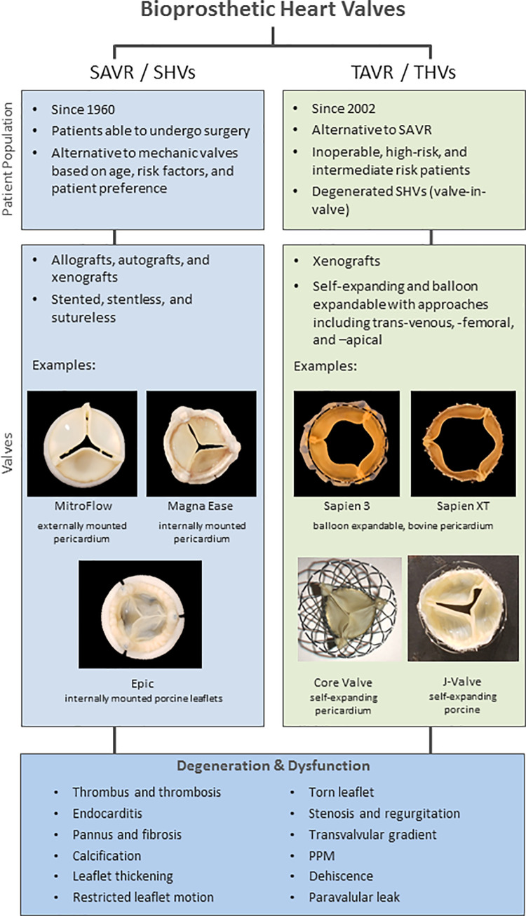
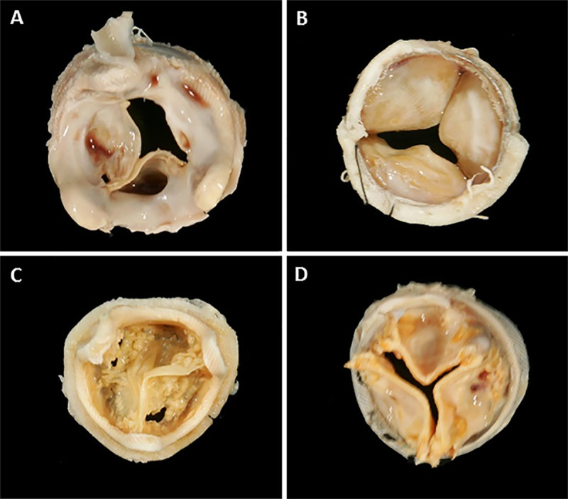
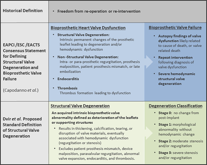
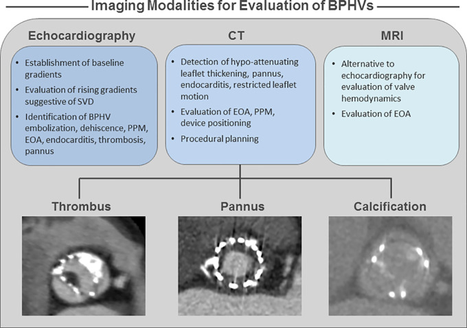
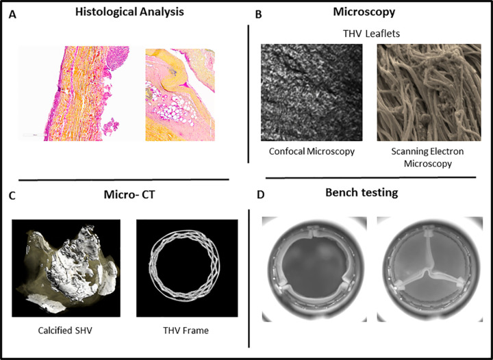
Bioprosthetic heart valves (BPHVs) have fundamentally changed the treatment of valvular heart disease. Despite the continuous progress of BPHVs, from early valve designs for use in surgical replacement to the rapidly evolving use of transcatheter replacement techniques and designs, valve dysfunction and degeneration remain fundamental issues. Current guidelines and proposed standard definitions of BPHV dysfunction and degeneration outline the importance of imaging. Imaging plays a key role in understanding valve degeneration, including clinical imaging to identify transvalvular gradients, leaflet thickening, thrombosis, calcification, and restricted or reduced leaflet motion. Similarly, translational imaging approaches-including micro-CT, high-speed video, computational modeling, and high-resolution microscopy-and histologic analysis are crucial to understanding mechanisms of valve degeneration and factors that may contribute to valve dysfunction. This article provides an overview of valve dysfunction and degeneration and the role of imaging. © RSNA, 2019.
2019 by the Radiological Society of North America, Inc.
Conflict of interest statement
Disclosures of Conflicts of Interest: S.L.S. disclosed no relevant relationships. P.B. Activities related to the present article: disclosed no relevant relationships. Activities not related to the present article: is a consultant for Circle Cardiovascular Imaging and has grants/grants pending with Abbott, Edwards Lifesciences, Medtronic, and Neovasc; institution received research support from Edwards. Other relationships: disclosed no relevant relationships. J.A.L. Activities related to the present article: disclosed no relevant relationships. Activities not related to the present article: is a consultant for Circle Cardiovascular Imaging and HeartFlow; institution has grants/grants pending with Abbott, Edwards Lifesciences, Medtronic, and Neovasc; institution received research support from Edwards. Other relationships: disclosed no relevant relationships.
Figures
References
-
- Iung B, Vahanian A. Epidemiology of acquired valvular heart disease. Can J Cardiol 2014;30(9):962–970. - PubMed
-
- Iung B, Vahanian A. Epidemiology of valvular heart disease in the adult. Nat Rev Cardiol 2011;8(3):162–172. - PubMed
-
- Borer J. The challenge: about the Heart Valve Society of America. Heart Valve Society of America. http://www.heartvalvesocietyofamerica.org/more.html. Published 2017. Accessed July 1, 2017.
Publication types
LinkOut - more resources
Full Text Sources
