

Radiomics for Distinguishing Myocardial Infarction from Myocarditis at Late Gadolinium Enhancement at MRI: Comparison with Subjective Visual Analysis
- PMID: 33778525
- PMCID: PMC7977789
- DOI: 10.1148/ryct.2019180026
Radiomics for Distinguishing Myocardial Infarction from Myocarditis at Late Gadolinium Enhancement at MRI: Comparison with Subjective Visual Analysis
Abstract
Purpose: To evaluate whether radiomics features of late gadolinium enhancement (LGE) regions at cardiac MRI enable distinction between myocardial infarction (MI) and myocarditis and to compare radiomics with subjective visual analyses by readers with different experience levels.
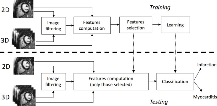
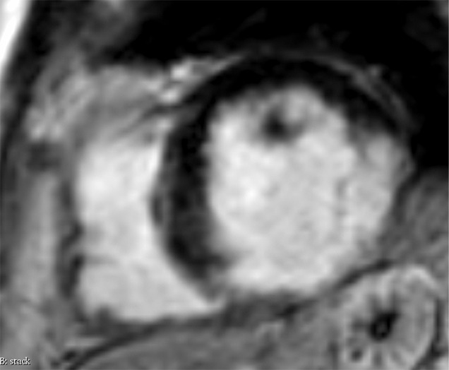
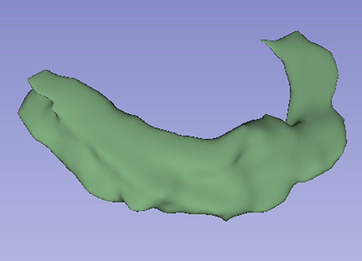
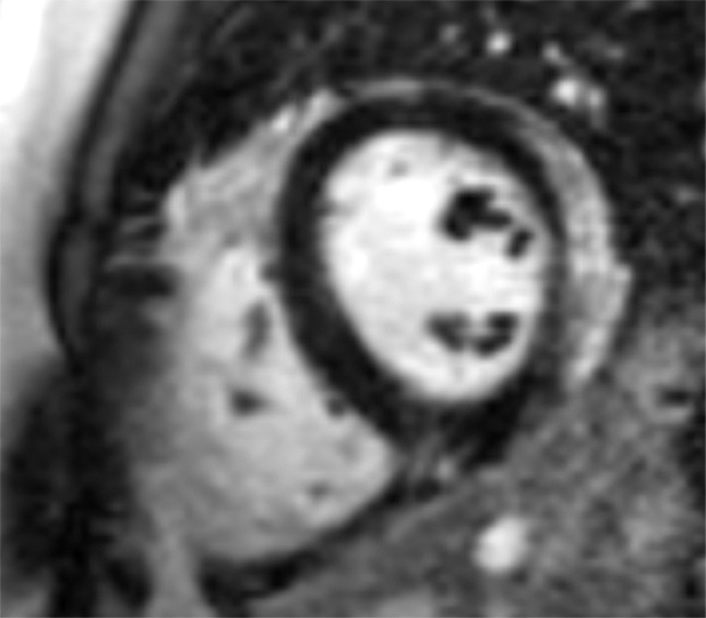
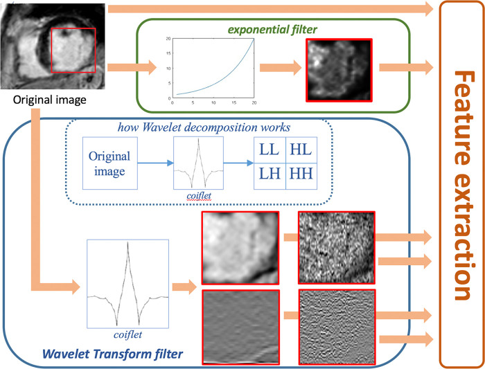
Materials and methods: In this retrospective, institutional review board-approved study, consecutive MRI examinations of 111 patients with MI and 62 patients with myocarditis showing LGE were included. By using open-source software, classification performances attained from two-dimensional (2D) and three-dimensional (3D) texture analysis, shape, and first-order descriptors were compared, applying five different machine learning algorithms. A nested, stratified 10-fold cross-validation was performed. Classification performances were compared through Wilcoxon signed-rank tests. Supervised and unsupervised feature selection techniques were tested; the effect of resampling MR images was analyzed. Subjective image analysis was performed on 2D and 3D image sets by two independent, blinded readers with different experience levels.
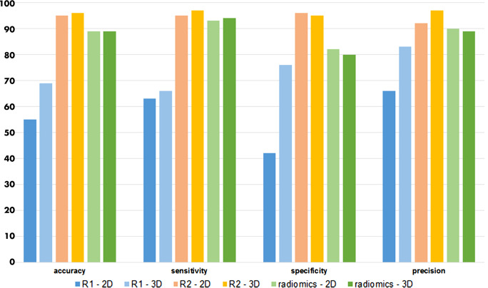
Results: When trained with recursive feature elimination (RFE), a support vector machine achieved the best results (accuracy: 88%) for 2D features, whereas linear discriminant analysis (LDA) showed the highest accuracy (85%) for 3D features (P <.05). When trained with principal component analysis (PCA), LDA attained the highest accuracy with both 2D (86%) and 3D (89%; P =.4) features. Results found for classifiers trained with spline resampling were less accurate than those achieved with one-dimensional (1D) nearest-neighbor interpolation (P <.05), whereas results for classifiers trained with 1D nearest-neighbor interpolation and without resampling were similar (P =.1). As compared with the radiomics approach, subjective visual analysis performance was lower for the less experienced and higher for the experienced reader for both 2D and 3D data.
Conclusion: Radiomics features of LGE permit the distinction between MI and myocarditis with high accuracy by using either 2D features and RFE or 3D features and PCA.© RSNA, 2019Supplemental material is available for this article.
2019 by the Radiological Society of North America, Inc.
Conflict of interest statement
Disclosures of Conflicts of Interest: T.D.N. Activities related to the present article: author received grant from European School of Radiology (research fellowship organized by ESOR in many European hospitals). Activities not related to the present article: disclosed no relevant relationships. Other relationships: disclosed no relevant relationships. J.v.S. disclosed no relevant relationships. M.M. disclosed no relevant relationships. E.G. disclosed no relevant relationships. P.S. disclosed no relevant relationships. R.M. disclosed no relevant relationships. H.A. disclosed no relevant relationships.
Figures

References
-
- van der Wall EE, Vliegen HW, de Roos A, Bruschke AV. Magnetic resonance imaging in coronary artery disease. Circulation 1995;92(9):2723–2739. - PubMed
-
- Berg J, Kottwitz J, Baltensperger N, et al. . Cardiac magnetic resonance imaging in myocarditis reveals persistent disease activity despite normalization of cardiac enzymes and inflammatory parameters at 3-month follow-up. Circ Heart Fail 2017;10(11):e004262. - PubMed
-
- Patriki D, Gresser E, Manka R, Emmert MY, Lüscher TF, Heidecker B. Approximation of the incidence of myocarditis by systematic screening with cardiac magnetic resonance imaging. JACC Heart Fail 2018;6(7):573–579. - PubMed
-
- Baessler B, Luecke C, Lurz J, et al. . Cardiac MRI texture analysis of T1 and T2 maps in patients with infarctlike acute myocarditis. Radiology 2018;289(2):357–365. - PubMed
LinkOut - more resources
Full Text Sources
Research Materials
