

Imaging Profile of the COVID-19 Infection: Radiologic Findings and Literature Review
- PMID: 33778547
- PMCID: PMC7233595
- DOI: 10.1148/ryct.2020200034
Imaging Profile of the COVID-19 Infection: Radiologic Findings and Literature Review
Abstract
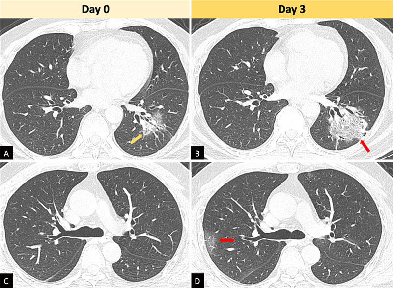
Purpose: To present the findings of 21 coronavirus disease 2019 (COVID-19) cases from two Chinese centers with CT and chest radiographic findings, as well as follow-up imaging in five cases.
Materials and methods: This was a retrospective study in Shenzhen and Hong Kong. Patients with COVID-19 infection were included. A systematic review of the published literature on radiologic features of COVID-19 infection was conducted.
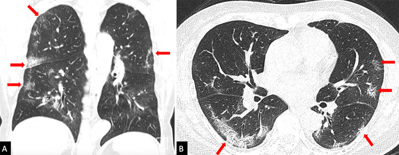
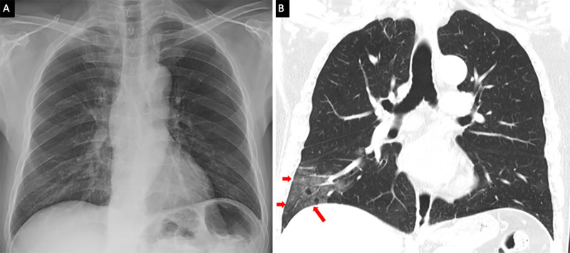
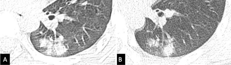
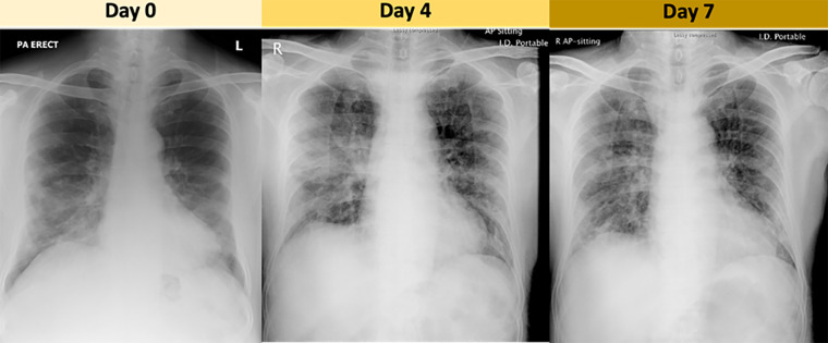
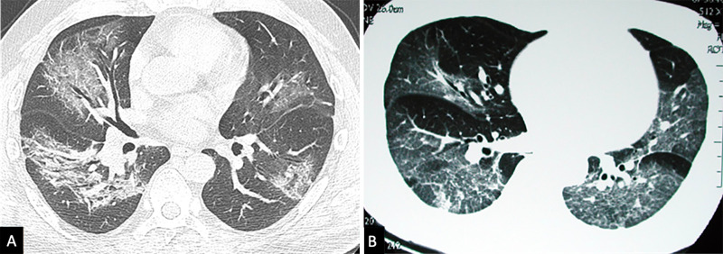
Results: The predominant imaging pattern was of ground-glass opacification with occasional consolidation in the peripheries. Pleural effusions and lymphadenopathy were absent in all cases. Patients demonstrated evolution of the ground-glass opacities into consolidation and subsequent resolution of the airspace changes. Ground-glass and consolidative opacities visible on CT are sometimes undetectable on chest radiography, suggesting that CT is a more sensitive imaging modality for investigation. The systematic review identified four other studies confirming the findings of bilateral and peripheral ground glass with or without consolidation as the predominant finding at CT chest examinations.
Conclusion: Pulmonary manifestation of COVID-19 infection is predominantly characterized by ground-glass opacification with occasional consolidation on CT. Radiographic findings in patients presenting in Shenzhen and Hong Kong are in keeping with four previous publications from other sites.© RSNA, 2020See editorial by Kay and Abbara in this issue.
2020 by the Radiological Society of North America, Inc.
Conflict of interest statement
Disclosures of Conflicts of Interest: : M.Y.G. disclosed no relevant relationships. E.Y.P.L. disclosed no relevant relationships. J.Y. disclosed no relevant relationships. F.Y. disclosed no relevant relationships. X.I. disclosed no relevant relationships. H.W. disclosed no relevant relationships. M.M.L. disclosed no relevant relationships. C.S.Y.L. disclosed no relevant relationships. B.L. disclosed no relevant relationships. P.L.K. disclosed no relevant relationships. C.K.M.H. disclosed no relevant relationships. K.Y. disclosed no relevant relationships. M.D.K. disclosed no relevant relationships.
Figures
Comment in
-
The Many Faces of COVID-19: Spectrum of Imaging Manifestations.Radiol Cardiothorac Imaging. 2020 Feb 14;2(1):e200037. doi: 10.1148/ryct.2020200037. eCollection 2020 Feb. Radiol Cardiothorac Imaging. 2020. PMID: 33779634 Free PMC article. No abstract available.
References
-
- World Health Organization . Novel Coronavirus(2019-nCoV) Situation Report - 11. https://www.who.int/docs/default-source/coronaviruse/situation-reports/2.... Published January 31, 2020.
-
- World Health Organization . Novel Coronavirus(2019-nCoV) Situation Report - 17. https://www.who.int/docs/default-source/coronaviruse/situation-reports/2.... Published February 6, 2020.
-
- Hansell DM, Bankier AA, MacMahon H, McLoud TC, Müller NL, Remy J. Fleischner Society: glossary of terms for thoracic imaging. Radiology 2008;246(3):697–722. - PubMed
LinkOut - more resources
Full Text Sources
