Multimodal Multiparametric Three-dimensional Image Fusion in Coronary Artery Disease: Combining the Best of Two Worlds
- PMID: 33778554
- PMCID: PMC7977970
- DOI: 10.1148/ryct.2020190116
Multimodal Multiparametric Three-dimensional Image Fusion in Coronary Artery Disease: Combining the Best of Two Worlds
Abstract
Purpose: To allow for comprehensive noninvasive diagnostics of coronary artery disease (CAD) by using three-dimensional (3D) image fusion of CT coronary angiography, CT-derived fractional flow reserve (CT FFR), whole-heart dynamic 3D cardiac MRI perfusion, and 3D cardiac MRI late gadolinium enhancement (LGE).
Materials and methods: Seventeen patients (54 years ± 10 [standard deviation], one female) who underwent cardiac CT and cardiac MRI were included (combined subcohort of three prospective trials). Software facilitating multimodal 3D image fusion was developed. Postprocessing of CT data included segmentation of the coronary tree and heart contours, calculation of CT FFR values, and color coding of the coronary tree according to CT FFR. Postprocessing of cardiac MRI data included segmentation of the left ventricle (LV) in cardiac MRI perfusion and cardiac MRI LGE, co-registration of cardiac MRI to CT data, and projection of cardiac MRI perfusion and LGE values onto the high spatial resolution LV from CT.
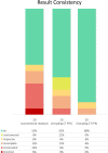
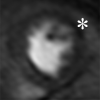
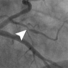
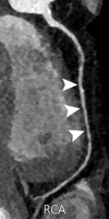
Results: Image quality was rated as good to excellent (scores: 2.5-2.6; 3 = excellent). CT coronary angiography revealed significant stenoses in seven of 17 cases (41%). CT FFR was possible in 16 of 17 cases (94%) and showed pathologic flow in seven of 17 cases (41%), six of which coincided with cases revealing significant stenoses at CT coronary angiography. Cardiac MRI perfusion identified eight of 17 patients (47%) with hypoperfusion (ischemic burden of 17% ± 5). Cardiac MRI LGE showed myocardial scar in three of 17 cases (18%, scar burden of 7% ± 4). Conventional two-dimensional readout of CT coronary angiography and cardiac MRI resulted in eight of 17 cases (47%) with uncertain findings. Most of these divergent findings could be solved when adding information from CT FFR and 3D image fusion (six of eight, 75%).
Conclusion: Multimodal 3D cardiac image fusion is feasible and may help with comprehensive noninvasive CAD diagnostics.Supplemental material is available for this article.© RSNA, 2020.
2020 by the Radiological Society of North America, Inc.
Conflict of interest statement
Disclosures of Conflicts of Interest: J.v.S. disclosed no relevant relationships. M.M. disclosed no relevant relationships. H.M. disclosed no relevant relationships. C.S. Activities related to the present article: disclosed no relevant relationships. Activities not related to the present article: is employed by and holds stock/stock options in Siemens Healthcare. Other relationships: has patents (pending and issued) with Siemens Healthcare. S.K. disclosed no relevant relationships. F.R. Activities related to the present article: disclosed no relevant relationships. Activities not related to the present article: is a board member of European Society of Cardiology; is a consultant for Amgen, Fresenius, CITI Research, and Vifor; has grants/grants pending with Abbott/St. Jude Medical, Amgen, Bayer, Novartis, Servier, and Swiss National Foundation; received payment for lectures including service on speakers bureaus from Abbott, AstraZeneca, Boehringer Ingelheim, Hôpitaux Universitaires des Genève/GECORE, Luzerner Kantonsspital, CCE Services (Boston Scientific), Medtronic, Medscape, Novartis, Roche, Ruwag, Sanofi-Aventis, Servier, and Swiss Heart Failure Academy (USZ). Other relationships: disclosed no relevant relationships. H.A. disclosed no relevant relationships. R.M. disclosed no relevant relationships.
Figures






References
-
- Patel MR, Calhoon JH, Dehmer GJ, et al. ACC/AATS/AHA/ASE/ASNC/SCAI/SCCT/STS 2017 appropriate use criteria for coronary revascularization in patients with stable ischemic heart disease: a report of the American College of Cardiology appropriate use criteria task force, American Association for Thoracic Surgery, American Heart Association, American Society of Echocardiography, American Society of Nuclear Cardiology, Society for Cardiovascular Angiography and Interventions, Society of Cardiovascular Computed Tomography, and Society of Thoracic Surgeons. [Published correction appears in J Am Coll Cardiol 2018;71(19):2279–2280]. J Am Coll Cardiol 2017;69(17):2212–2241. - PubMed
-
- Hausleiter J, Meyer T, Hadamitzky M, et al. Non-invasive coronary computed tomographic angiography for patients with suspected coronary artery disease: the Coronary Angiography by Computed Tomography with the Use of a Submillimeter resolution (CACTUS) trial. Eur Heart J 2007;28(24):3034–3041. - PubMed
-
- Itu L, Rapaka S, Passerini T, et al. A machine-learning approach for computation of fractional flow reserve from coronary computed tomography. J Appl Physiol (1985) 2016;121(1):42–52. - PubMed
-
- Tesche C, De Cecco CN, Albrecht MH, et al. Coronary CT angiography-derived fractional flow reserve. Radiology 2017;285(1):17–33. - PubMed
-
- Ihdayhid AR, Norgaard BL, Gaur S, et al. Prognostic value and risk continuum of noninvasive fractional flow reserve derived from coronary CT angiography. Radiology 2019;292(2):343–351. - PubMed
LinkOut - more resources
Full Text Sources
Miscellaneous

