Chest CT Severity Score: An Imaging Tool for Assessing Severe COVID-19
- PMID: 33778560
- PMCID: PMC7233443
- DOI: 10.1148/ryct.2020200047
Chest CT Severity Score: An Imaging Tool for Assessing Severe COVID-19
Abstract
Purpose: To evaluate the value of chest CT severity score (CT-SS) in differentiating clinical forms of coronavirus disease 2019 (COVID-19).
Materials and methods: A total of 102 patients with COVID-19 confirmed by a positive result from real-time reverse transcription polymerase chain reaction on throat swabs who underwent chest CT (53 men and 49 women, 15-79 years old, 84 cases with mild and 18 cases with severe disease) were included in the study. The CT-SS was defined by summing up individual scores from 20 lung regions; scores of 0, 1, and 2 were respectively assigned for each region if parenchymal opacification involved 0%, less than 50%, or equal to or more than 50% of each region (theoretic range of CT-SS from 0 to 40). The clinical and laboratory data were collected, and patients were clinically subdivided according to disease severity according to the Chinese National Health Commission guidelines.
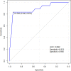
Results: The posterior segment of upper lobe (left, 68 of 102; right, 68 of 102), superior segment of lower lobe (left, 79 of 102; right, 79 of 102), lateral basal segment (left, 79 of 102; right, 70 of 102), and posterior basal segment of lower lobe (left, 81 of 102; right, 83 of 102) were the most frequently involved sites in COVID-19. Lung opacification mainly involved the lower lobes, in comparison with middle-upper lobes. No significant differences in distribution of the disease were seen between right and left lungs. The individual scores in each lung and the total CT-SS were higher in severe COVID-19 when compared with mild cases (P < .05). The optimal CT-SS threshold for identifying severe COVID-19 was 19.5 (area under curve = 0.892), with 83.3% sensitivity and 94% specificity.
Conclusion: The CT-SS could be used to evaluate the severity of pulmonary involvement quickly and objectively in patients with COVID-19.© RSNA, 2020.
2020 by the Radiological Society of North America, Inc.
Conflict of interest statement
Disclosures of Conflicts of Interest: R.Y. disclosed no relevant relationships. X.L. disclosed no relevant relationships. H.L. disclosed no relevant relationships. Y.Z. disclosed no relevant relationships. X.Z. disclosed no relevant relationships. Q.X. disclosed no relevant relationships. Y.L. disclosed no relevant relationships. C.G. disclosed no relevant relationships. W.Z. disclosed no relevant relationships.
Figures


References
-
- Gorbalenya AE, Baker SC, Baric RS, et al. Severe acute respiratory syndrome-related coronavirus: The species and its viruses – a statement of the Coronavirus Study Group. bioRxiv [preprint] https://www.biorxiv.org/content/10.1101/2020.02.07.937862v1. - DOI
-
- World Health Organization. Novel coronavirus – China. Feb 11, 2020. https://www.who.int/docs/default-source/coronaviruse/situation-reports/2....
-
- Zhou P, Yang XL, Wang XG, et al. Discovery of a novel coronavirus associated with the recent pneumonia outbreak in humans and its potential bat origin. bioRxiv [preprint]. https://www.biorxiv.org/content/10.1101/2020.01.22.914952v2. - DOI
LinkOut - more resources
Full Text Sources
Miscellaneous

