. 2020 Mar 17;2(2):e200110.
doi: 10.1148/ryct.2020200110. eCollection 2020 Apr.

Chest CT Findings in Cases from the Cruise Ship Diamond Princess with Coronavirus Disease (COVID-19)

Affiliations

Chest CT Findings in Cases from the Cruise Ship Diamond Princess with Coronavirus Disease (COVID-19)

Shohei Inui et al. Radiol Cardiothorac Imaging. .

Erratum in

Abstract

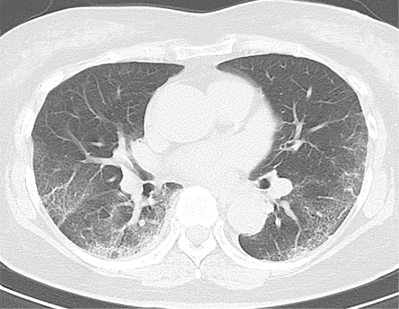
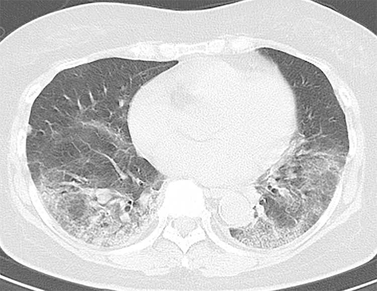
Purpose: To evaluate the chest CT findings in an environmentally homogeneous cohort from the cruise ship Diamond Princess with coronavirus disease 2019 (COVID-19).

Materials and methods: This retrospective study comprised 104 cases (mean age, 62 years ± 16 [standard deviation], range, 25-93 years) with COVID-19 confirmed with reverse-transcription polymerase change reaction findings. CT images were reviewed, and the CT severity score was calculated for each lobe and the entire lung. CT findings were compared between asymptomatic and symptomatic cases.

Results: Of 104 cases, 76 (73%) were asymptomatic, 41 (54%) of which had lung opacities on CT. Twenty-eight (27%) cases were symptomatic, 22 (79%) of which had abnormal CT findings. Symptomatic cases showed lung opacities and airway abnormalities on CT more frequently than asymptomatic cases [lung opacity; 22 (79%) vs 41 (54%), airway abnormalities; 14 (50%) vs 15 (20%)]. Asymptomatic cases showed more ground-glass opacity (GGO) over consolidation (83%), while symptomatic cases more frequently showed consolidation over GGO (41%). The CT severity score was higher in symptomatic cases than asymptomatic cases, particularly in the lower lobes [symptomatic vs asymptomatic cases; right lower lobe: 2 ± 1 (0-4) vs 1 ± 1 (0-4); left lower lobe: 2 ± 1 (0-4) vs 1 ± 1 (0-3); total score: 7 ± 5 (1-17) vs 4 ± 2 (1-11)].

Conclusion: This study documented a high incidence of subclinical CT changes in cases with COVID-19. Compared with symptomatic cases, asymptomatic cases showed more GGO over consolidation and milder extension of disease on CT.An earlier incorrect version appeared online. This article was corrected on April 8, 2020.© RSNA, 2020.


Conflict of interest statement

Disclosures of Conflicts of Interest: S.I. disclosed no relevant relationships. A.F. disclosed no relevant relationships. M.J. disclosed no relevant relationships. N.K. disclosed no relevant relationships. S.W. disclosed no relevant relationships. Y.S. disclosed no relevant relationships. S.U. disclosed no relevant relationships. Y.U. disclosed no relevant relationships.

Figures

A 70-year-old asymptomatic woman. On axial CT image, focal subpleural ground-glass opacities with smooth intralobular and interlobular septal thickening were demonstrated in the right and left lower lobes (arrows). The left lower lobe lesion was accompanied by air bronchogram with mild bronchial dilatation (arrowhead).
Figure 1:
A 70-year-old asymptomatic woman. On axial CT image, focal subpleural ground-glass opacities with smooth intralobular and interlobular septal thickening were demonstrated in the right and left lower lobes (arrows). The left lower lobe lesion was accompanied by air bronchogram with mild bronchial dilatation (arrowhead).
A 66-year-old asymptomatic woman. On axial CT images, focal rounded ground-glass opacities (arrows) with partial consolidation in a peribronchial and subpleural distribution were noted in the right upper (a), middle (b), and lower (c) lobes and left lower (b) lobe.
Figure 2a:
A 66-year-old asymptomatic woman. On axial CT images, focal rounded ground-glass opacities (arrows) with partial consolidation in a peribronchial and subpleural distribution were noted in the right upper (a), middle (b), and lower (c) lobes and left lower (b) lobe.
Figure 2b:
A 66-year-old asymptomatic woman. On axial CT images, focal rounded ground-glass opacities (arrows) with partial consolidation in a peribronchial and subpleural distribution were noted in the right upper (a), middle (b), and lower (c) lobes and left lower (b) lobe.
Figure 2c:
A 66-year-old asymptomatic woman. On axial CT images, focal rounded ground-glass opacities (arrows) with partial consolidation in a peribronchial and subpleural distribution were noted in the right upper (a), middle (b), and lower (c) lobes and left lower (b) lobe.
A 73-year-old asymptomatic woman. (a) On axial CT images, focal peripheral ground-glass opacities with intralobular and interlobular smooth septal thickening were shown in the left (arrowhead) and right upper lobe (arrows). The right upper lobe lesions were accompanied by subpleural curvilinear lines (arrow). (b, c) Diffuse ground-glass (reticular) opacities with consolidation with bronchiectasis and bronchial wall thickening were demonstrated in the left and right lower lobes.
Figure 3a:
A 73-year-old asymptomatic woman. (a) On axial CT images, focal peripheral ground-glass opacities with intralobular and interlobular smooth septal thickening were shown in the left (arrowhead) and right upper lobe (arrows). The right upper lobe lesions were accompanied by subpleural curvilinear lines (arrow). (b, c) Diffuse ground-glass (reticular) opacities with consolidation with bronchiectasis and bronchial wall thickening were demonstrated in the left and right lower lobes.
Figure 3b:
A 73-year-old asymptomatic woman. (a) On axial CT images, focal peripheral ground-glass opacities with intralobular and interlobular smooth septal thickening were shown in the left (arrowhead) and right upper lobe (arrows). The right upper lobe lesions were accompanied by subpleural curvilinear lines (arrow). (b, c) Diffuse ground-glass (reticular) opacities with consolidation with bronchiectasis and bronchial wall thickening were demonstrated in the left and right lower lobes.
Figure 3c:
A 73-year-old asymptomatic woman. (a) On axial CT images, focal peripheral ground-glass opacities with intralobular and interlobular smooth septal thickening were shown in the left (arrowhead) and right upper lobe (arrows). The right upper lobe lesions were accompanied by subpleural curvilinear lines (arrow). (b, c) Diffuse ground-glass (reticular) opacities with consolidation with bronchiectasis and bronchial wall thickening were demonstrated in the left and right lower lobes.

References

    1. World Health Organization . Naming the coronavirus disease (COVID-19) and the virus that causes it. https://www.who.int/emergencies/diseases/novel-coronavirus-2019/technica.... Accessed March 5, 2020.
    1. Huang C, Wang Y, Li X, et al. Clinical features of patients infected with 2019 novel coronavirus in Wuhan, China. Lancet 2020;395(10223):497–506 [Published correction appears in Lancet 2020;395(10223):496.]. - PMC - PubMed
    1. The Ministry of Health, Labour and Welfare . The infection control measures taken at the Cruise ship “Diamond Princess” (provisional translation). https://www.mhlw.go.jp/stf/seisakunitsuite/newpage_00001.html. Accessed March 5, 2020.
    1. Shi H, Han X, Jiang N, et al. Radiological findings from 81 patients with COVID-19 pneumonia in Wuhan, China: a descriptive study. Lancet Infect Dis 2020 Feb 24 [Epub ahead of print]. - PMC - PubMed
    1. Chung M, Bernheim A, Mei X, et al. CT imaging features of 2019 novel coronavirus (2019-nCoV). Radiology 2020;295(1):202–207. - PMC - PubMed

LinkOut - more resources