. 2020 Apr 6;2(2):e200117.
doi: 10.1148/ryct.2020200117. eCollection 2020 Apr.

Differences in Clinical and Imaging Presentation of Pediatric Patients with COVID-19 in Comparison with Adults

Affiliations

Differences in Clinical and Imaging Presentation of Pediatric Patients with COVID-19 in Comparison with Adults

Amei Chen et al. Radiol Cardiothorac Imaging. .

Abstract

Purpose: To characterize and compare the initial clinical and imaging features of coronavirus disease 2019 (COVID-19) in pediatric and adult patients undergoing chest CT.

Materials and methods: A total of 61 patients, consisting of 47 adults (aged 18 years or older) and 14 pediatric patients (aged younger than 18 years) with laboratory-confirmed COVID-19 confirmed by real-time reverse-transcription polymerase chain reaction between January 25 and February 15, 2020, were enrolled in this study. All patients underwent chest CT within 3 days after the initial reverse transcription polymerase chain reaction test. The clinical presentation, serum markers, and CT findings were assessed and compared between the adult and pediatric patients.

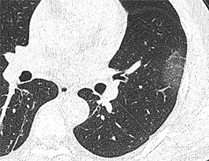
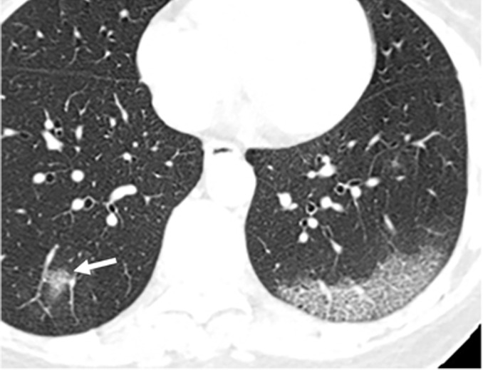
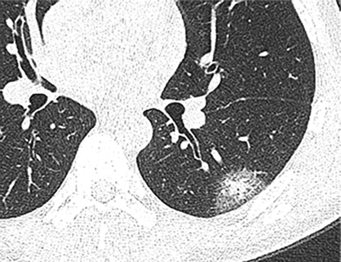
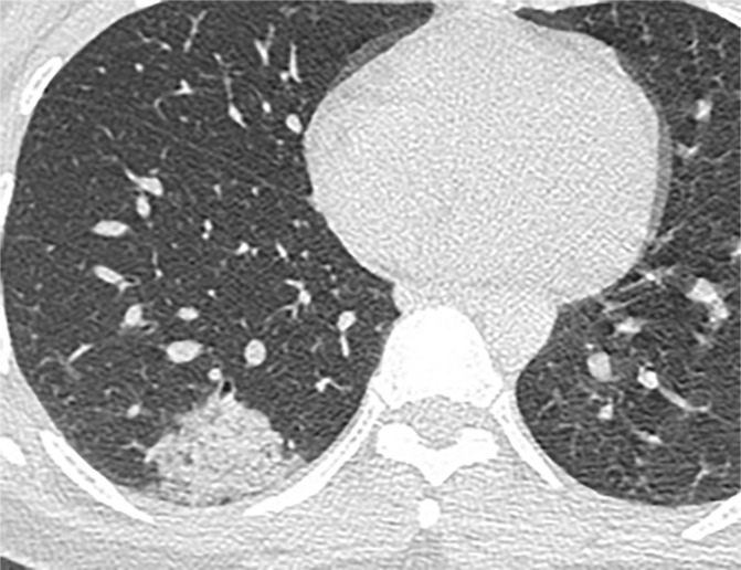
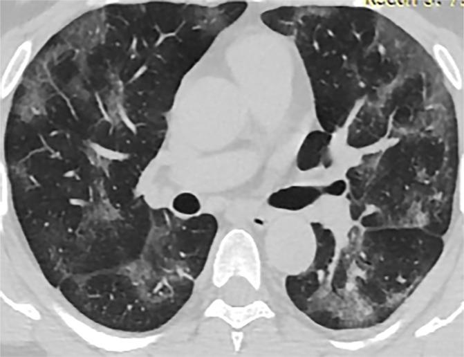
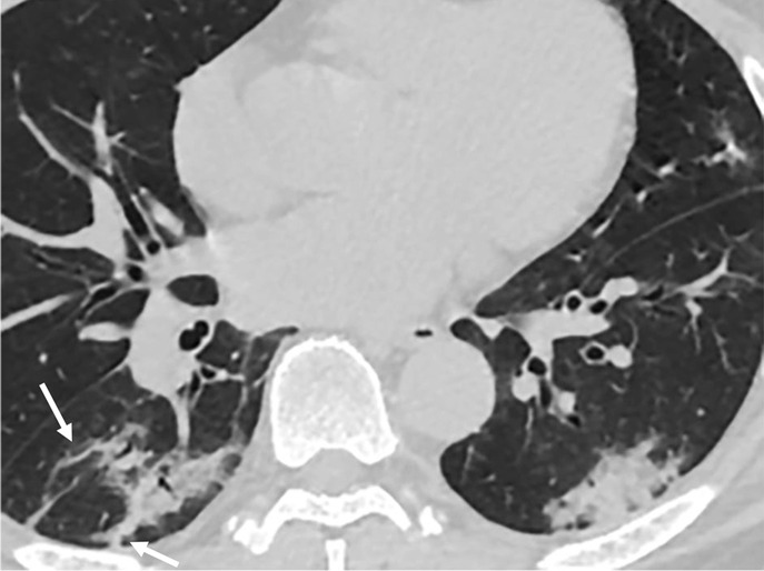
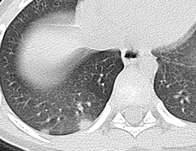
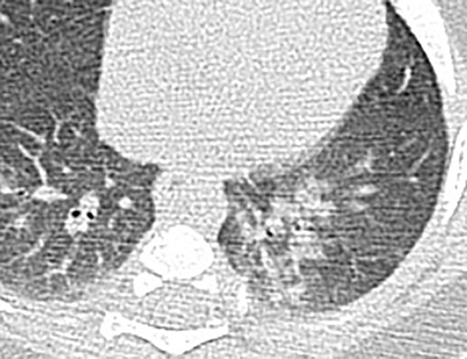
Results: Fever was less common in pediatric patients than in adults (six of 14, 42.9% vs 39 of 47, 83%; P = .008). Leukopenia or normal, lymphopenia or normal, and increased or normal C-reactive protein level were common in both groups with no difference (P > .05). Compared with the adults, pediatric patients had a lower rate of positive CT findings and a milder clinical grade (P = .004 and P = .001, respectively). At chest CT, the number of pulmonary lobes involved was found to be reduced in pediatric patients when compared with adults (P = .012). Subpleural distribution of lung opacities was a dominant feature in both groups, whereas bronchial distribution was more common in the pediatric group (P = .048). Among the CT features in adults, ground-glass opacities (GGOs) were the most common finding (24 of 43, 53.5%), followed by GGO with consolidation (14 of 43, 27.9%). In pediatric patients, GGOs accounted for 42.9% (three of seven), bronchial wall thickening occurred in 28.6% (two of seven), and GGOs with consolidations and nodular opacities occurred in 14.3% (one of seven). However, these CT features did not differ in the two groups, except for bronchial wall thickening, which was more commonly found in pediatric patients (P = .048). In addition, the semiquantitative scores of lung involvement were higher in adults than in pediatric patients (8.89 ± 4.54 vs 1.86 ± 2.41; P < .001).

Conclusion: Compared with adults, pediatric patients with COVID-19 showed distinctive clinical and CT features. Pediatric patients tend to have milder clinical symptoms, fewer positive results at CT, and less extensive involvement at imaging. Bronchial wall thickening was relatively more frequent on CT images from pediatric patients with COVID-19 in comparison with adults.Supplemental material is available for this article.© RSNA, 2020.


Conflict of interest statement

Disclosures of Conflicts of Interest: A.C. disclosed no relevant relationships. J.H. disclosed no relevant relationships. Y.L. disclosed no relevant relationships. Z.L. disclosed no relevant relationships. D.C. disclosed no relevant relationships. C.Y. disclosed no relevant relationships. R.Y. disclosed no relevant relationships. X.W. disclosed no relevant relationships.

Figures

Chest CT findings in adult patients with COVID-19 on transaxial images. (a) Woman, 36 years old, 1 day after symptom onset. Subpleural ground-glass opacity in left lower lobe. (b) Man, 54 years old, 4 days after symptom onset. Subpleural ground-glass opacity in left lower lobe with inter- and intralobular septal thickening (crazy paving) and a ground-glass nodule in the right lower lobe (arrow). (c) Man, 28 years old, 3 days after symptom onset. Subpleural ground-glass opacity in the left lower lobe with central consolidation. (d) Woman, 49 years old, 7 days after symptom onset. Pure consolidation in right lower lobe. (e) Man, 42 years old, 6 days after symptom onset. Bilateral multifocal pure ground-glass opacities. (f) Man, 62 years old, 14 days after symptom onset, bilateral foci of consolidation in both lower lobes, with early linear opacities in a perilobular pattern (arrows).
Figure 1a:
Chest CT findings in adult patients with COVID-19 on transaxial images. (a) Woman, 36 years old, 1 day after symptom onset. Subpleural ground-glass opacity in left lower lobe. (b) Man, 54 years old, 4 days after symptom onset. Subpleural ground-glass opacity in left lower lobe with inter- and intralobular septal thickening (crazy paving) and a ground-glass nodule in the right lower lobe (arrow). (c) Man, 28 years old, 3 days after symptom onset. Subpleural ground-glass opacity in the left lower lobe with central consolidation. (d) Woman, 49 years old, 7 days after symptom onset. Pure consolidation in right lower lobe. (e) Man, 42 years old, 6 days after symptom onset. Bilateral multifocal pure ground-glass opacities. (f) Man, 62 years old, 14 days after symptom onset, bilateral foci of consolidation in both lower lobes, with early linear opacities in a perilobular pattern (arrows).
Figure 1b:
Chest CT findings in adult patients with COVID-19 on transaxial images. (a) Woman, 36 years old, 1 day after symptom onset. Subpleural ground-glass opacity in left lower lobe. (b) Man, 54 years old, 4 days after symptom onset. Subpleural ground-glass opacity in left lower lobe with inter- and intralobular septal thickening (crazy paving) and a ground-glass nodule in the right lower lobe (arrow). (c) Man, 28 years old, 3 days after symptom onset. Subpleural ground-glass opacity in the left lower lobe with central consolidation. (d) Woman, 49 years old, 7 days after symptom onset. Pure consolidation in right lower lobe. (e) Man, 42 years old, 6 days after symptom onset. Bilateral multifocal pure ground-glass opacities. (f) Man, 62 years old, 14 days after symptom onset, bilateral foci of consolidation in both lower lobes, with early linear opacities in a perilobular pattern (arrows).
Figure 1c:
Chest CT findings in adult patients with COVID-19 on transaxial images. (a) Woman, 36 years old, 1 day after symptom onset. Subpleural ground-glass opacity in left lower lobe. (b) Man, 54 years old, 4 days after symptom onset. Subpleural ground-glass opacity in left lower lobe with inter- and intralobular septal thickening (crazy paving) and a ground-glass nodule in the right lower lobe (arrow). (c) Man, 28 years old, 3 days after symptom onset. Subpleural ground-glass opacity in the left lower lobe with central consolidation. (d) Woman, 49 years old, 7 days after symptom onset. Pure consolidation in right lower lobe. (e) Man, 42 years old, 6 days after symptom onset. Bilateral multifocal pure ground-glass opacities. (f) Man, 62 years old, 14 days after symptom onset, bilateral foci of consolidation in both lower lobes, with early linear opacities in a perilobular pattern (arrows).
Figure 1d:
Chest CT findings in adult patients with COVID-19 on transaxial images. (a) Woman, 36 years old, 1 day after symptom onset. Subpleural ground-glass opacity in left lower lobe. (b) Man, 54 years old, 4 days after symptom onset. Subpleural ground-glass opacity in left lower lobe with inter- and intralobular septal thickening (crazy paving) and a ground-glass nodule in the right lower lobe (arrow). (c) Man, 28 years old, 3 days after symptom onset. Subpleural ground-glass opacity in the left lower lobe with central consolidation. (d) Woman, 49 years old, 7 days after symptom onset. Pure consolidation in right lower lobe. (e) Man, 42 years old, 6 days after symptom onset. Bilateral multifocal pure ground-glass opacities. (f) Man, 62 years old, 14 days after symptom onset, bilateral foci of consolidation in both lower lobes, with early linear opacities in a perilobular pattern (arrows).
Figure 1e:
Chest CT findings in adult patients with COVID-19 on transaxial images. (a) Woman, 36 years old, 1 day after symptom onset. Subpleural ground-glass opacity in left lower lobe. (b) Man, 54 years old, 4 days after symptom onset. Subpleural ground-glass opacity in left lower lobe with inter- and intralobular septal thickening (crazy paving) and a ground-glass nodule in the right lower lobe (arrow). (c) Man, 28 years old, 3 days after symptom onset. Subpleural ground-glass opacity in the left lower lobe with central consolidation. (d) Woman, 49 years old, 7 days after symptom onset. Pure consolidation in right lower lobe. (e) Man, 42 years old, 6 days after symptom onset. Bilateral multifocal pure ground-glass opacities. (f) Man, 62 years old, 14 days after symptom onset, bilateral foci of consolidation in both lower lobes, with early linear opacities in a perilobular pattern (arrows).
Figure 1f:
Chest CT findings in adult patients with COVID-19 on transaxial images. (a) Woman, 36 years old, 1 day after symptom onset. Subpleural ground-glass opacity in left lower lobe. (b) Man, 54 years old, 4 days after symptom onset. Subpleural ground-glass opacity in left lower lobe with inter- and intralobular septal thickening (crazy paving) and a ground-glass nodule in the right lower lobe (arrow). (c) Man, 28 years old, 3 days after symptom onset. Subpleural ground-glass opacity in the left lower lobe with central consolidation. (d) Woman, 49 years old, 7 days after symptom onset. Pure consolidation in right lower lobe. (e) Man, 42 years old, 6 days after symptom onset. Bilateral multifocal pure ground-glass opacities. (f) Man, 62 years old, 14 days after symptom onset, bilateral foci of consolidation in both lower lobes, with early linear opacities in a perilobular pattern (arrows).
Chest CT findings of pediatric patients with COVID-19 on transaxial images. (a) Male patient, 2 months old, 2 days after symptom onset. Patchy ground-glass opacities (GGOs) in the right lower lobe. (b) Female patient, 4 years old, 4 days after symptom onset, two subpleural nodules in the right lower lobe. (c) Male patient, 8 months old, 6 days after symptom onset. Bronchial wall thickening and peribronchial GGOs and consolidation are noted in the left lower lobe.
Figure 2a:
Chest CT findings of pediatric patients with COVID-19 on transaxial images. (a) Male patient, 2 months old, 2 days after symptom onset. Patchy ground-glass opacities (GGOs) in the right lower lobe. (b) Female patient, 4 years old, 4 days after symptom onset, two subpleural nodules in the right lower lobe. (c) Male patient, 8 months old, 6 days after symptom onset. Bronchial wall thickening and peribronchial GGOs and consolidation are noted in the left lower lobe.
Figure 2b:
Chest CT findings of pediatric patients with COVID-19 on transaxial images. (a) Male patient, 2 months old, 2 days after symptom onset. Patchy ground-glass opacities (GGOs) in the right lower lobe. (b) Female patient, 4 years old, 4 days after symptom onset, two subpleural nodules in the right lower lobe. (c) Male patient, 8 months old, 6 days after symptom onset. Bronchial wall thickening and peribronchial GGOs and consolidation are noted in the left lower lobe.
Figure 2c:
Chest CT findings of pediatric patients with COVID-19 on transaxial images. (a) Male patient, 2 months old, 2 days after symptom onset. Patchy ground-glass opacities (GGOs) in the right lower lobe. (b) Female patient, 4 years old, 4 days after symptom onset, two subpleural nodules in the right lower lobe. (c) Male patient, 8 months old, 6 days after symptom onset. Bronchial wall thickening and peribronchial GGOs and consolidation are noted in the left lower lobe.

Similar articles

Cited by

References

    1. World Health Organization . Novel coronavirus – China. http://www.who.int/csr/don/12-january-2020-novel-coronavirus-china/en/. Published January 12, 2020. Accessed January 19, 2020.
    1. World Health Organization . Novel coronavirus – Thailand (ex-China). http://www.who.int/csr/don/14-january-2020-novel-coronavirus-thailand/en/. Published January 14, 2020. Accessed January 19, 2020.
    1. World Health Organization . Novel coronavirus – Japan (ex-China). http://www.who.int/csr/don/17-january-2020-novel-coronavirus-japan-ex-ch.... Published January 17, 2020. Accessed January 19, 2020.
    1. World Health Organization . Novel coronavirus – Republic of Korea (ex-China). http://www.who.int/csr/don/21-january-2020-novel-coronavirus-republic-of.... Published January 21, 2020. Accessed January 23, 2020.
    1. Ksiazek TG, Erdman D, Goldsmith CS, et al. A novel coronavirus associated with severe acute respiratory syndrome. N Engl J Med 2003;348(20):1953–1966. - PubMed