. 2020 Apr 20;2(2):e200130.
doi: 10.1148/ryct.2020200130. eCollection 2020 Apr.

CT Features and Short-term Prognosis of COVID-19 Pneumonia: A Single-Center Study from Kashan, Iran

Affiliations

CT Features and Short-term Prognosis of COVID-19 Pneumonia: A Single-Center Study from Kashan, Iran

Seyed Mohammad Hossein Tabatabaei et al. Radiol Cardiothorac Imaging. .

Abstract

Purpose: To assess whether certain CT chest features of patients with confirmed coronavirus disease 2019 (COVID-19) may have short-term prognostic value.

Materials and methods: One hundred-twenty consecutive symptomatic patients with COVID-19 infection who had undergone chest CT were enrolled in this retrospective study. Patients were categorized into three groups: routine inward hospitalization, intensive care unit admission, and deceased based on a short-term follow-up. Detailed initial CT features and distributional evaluation were recorded.

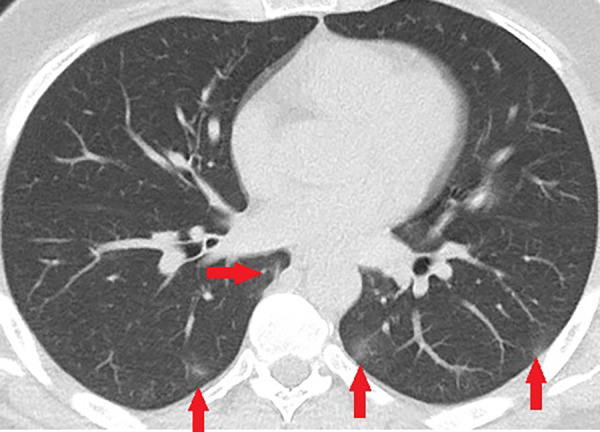
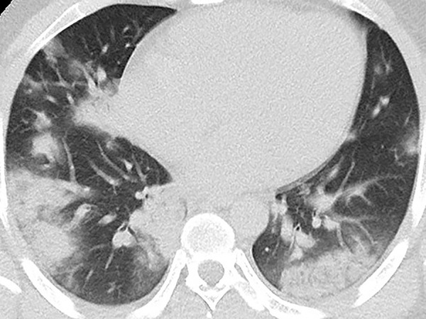
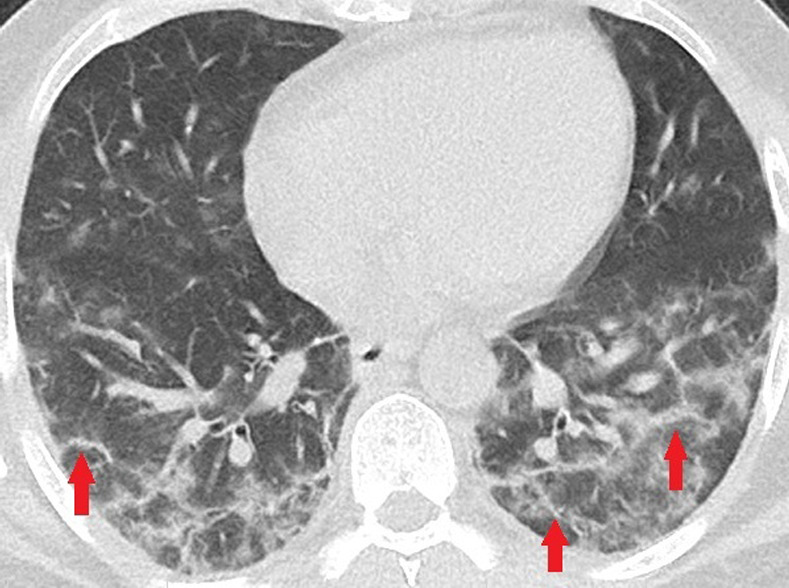
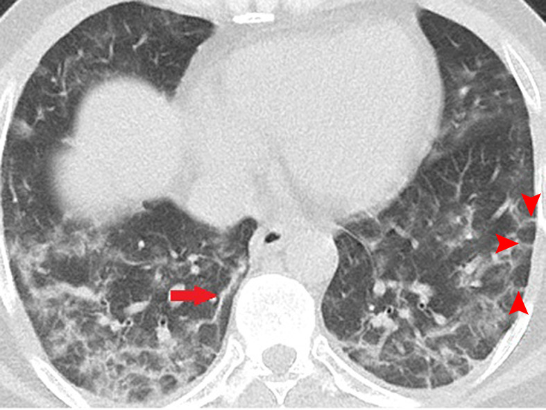
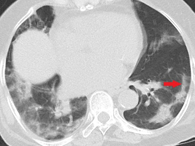
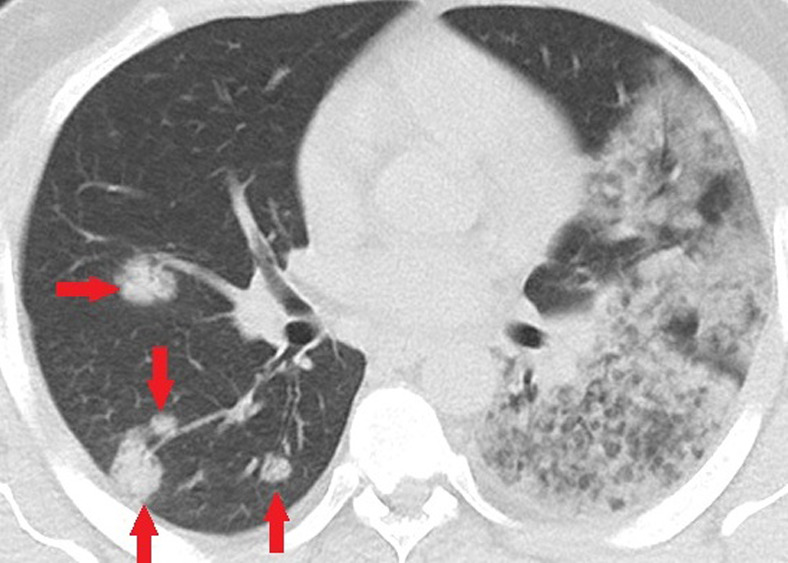
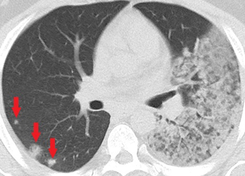
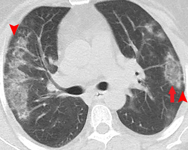
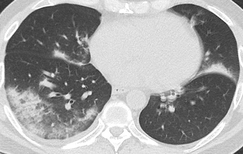
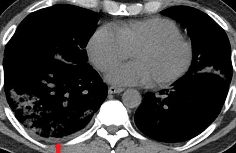
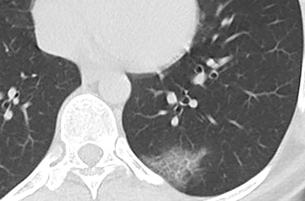
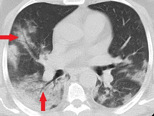
Results: The mean age in the deceased group was 70.7 years, significantly higher than the other two groups (P < .05). Ninety-four percent (113/120) of the patients had ground-glass opacities (GGO). Peripheral and lower zone predilection was present in most patients. Subpleural sparing and pleural effusion were seen in approximately 23% (28/120) and 17% (20/120) of the patients, respectively. The combined intensive care unit group and deceased patients had significantly more consolidation, air bronchograms, crazy paving, and central involvement of the lungs compared with routinely hospitalized patients (all P < .05).

Conclusion: This study supports the previously described typical CT appearance of COVID-19 pneumonia with bilateral GGO, in peripheral distribution and lower lung zone predilection. Subpleural sparing and pleural effusion were seen approximately in one-fifth and one-sixth of the patients with COVID-19, respectively. Consolidation, air bronchograms, central lung involvement, crazy paving and pleural effusion on initial CT chest have potential prognostic values, the features more commonly observed in critically ill patients.© RSNA, 2020.


Conflict of interest statement

Disclosures of Conflicts of Interest: S.M.H.T. disclosed no relevant relationships. H.T. disclosed no relevant relationships. F.M. disclosed no relevant relationships. H.R. disclosed no relevant relationships.

Figures

Unenhanced CT of the chest of a 43-year-old man with confirmed COVID-19 pneumonia. Few small peripheral patchy ground-glass opacities (arrows) are the only positive findings on the chest CT of this patient.
Figure 1:
Unenhanced CT of the chest of a 43-year-old man with confirmed COVID-19 pneumonia. Few small peripheral patchy ground-glass opacities (arrows) are the only positive findings on the chest CT of this patient.
Unenhanced CT of the chest of a 28-year-old man with confirmed COVID-19 pneumonia. Multiple peripheral consolidations are shown bilaterally, with patchy and segmental morphology.
Figure 2:
Unenhanced CT of the chest of a 28-year-old man with confirmed COVID-19 pneumonia. Multiple peripheral consolidations are shown bilaterally, with patchy and segmental morphology.
Unenhanced CT of the chest of two patients with confirmed COVID-19 pneumonia. (a, b) Multiple bilateral linear and reticular opacities (arrows) in a 50-year-old man. Note the linear opacities with an arc pattern (arrowheads) on the left side, representing perilobular opacities, called perilobular sign which is a typical sign of organizing pneumonia. Note some of the mentioned opacities on b outline borders of secondary pulmonary lobules (arrowheads), representing interstitial pneumonia. (c) A peripheral linear consolidation (arrow) in the left lung as well as ground-glass opacities and consolidations in some other parts of both lungs in an 81-year-old woman.
Figure 3a:
Unenhanced CT of the chest of two patients with confirmed COVID-19 pneumonia. (a, b) Multiple bilateral linear and reticular opacities (arrows) in a 50-year-old man. Note the linear opacities with an arc pattern (arrowheads) on the left side, representing perilobular opacities, called perilobular sign which is a typical sign of organizing pneumonia. Note some of the mentioned opacities on b outline borders of secondary pulmonary lobules (arrowheads), representing interstitial pneumonia. (c) A peripheral linear consolidation (arrow) in the left lung as well as ground-glass opacities and consolidations in some other parts of both lungs in an 81-year-old woman.
Figure 3b:
Unenhanced CT of the chest of two patients with confirmed COVID-19 pneumonia. (a, b) Multiple bilateral linear and reticular opacities (arrows) in a 50-year-old man. Note the linear opacities with an arc pattern (arrowheads) on the left side, representing perilobular opacities, called perilobular sign which is a typical sign of organizing pneumonia. Note some of the mentioned opacities on b outline borders of secondary pulmonary lobules (arrowheads), representing interstitial pneumonia. (c) A peripheral linear consolidation (arrow) in the left lung as well as ground-glass opacities and consolidations in some other parts of both lungs in an 81-year-old woman.
Figure 3c:
Unenhanced CT of the chest of two patients with confirmed COVID-19 pneumonia. (a, b) Multiple bilateral linear and reticular opacities (arrows) in a 50-year-old man. Note the linear opacities with an arc pattern (arrowheads) on the left side, representing perilobular opacities, called perilobular sign which is a typical sign of organizing pneumonia. Note some of the mentioned opacities on b outline borders of secondary pulmonary lobules (arrowheads), representing interstitial pneumonia. (c) A peripheral linear consolidation (arrow) in the left lung as well as ground-glass opacities and consolidations in some other parts of both lungs in an 81-year-old woman.
Unenhanced CT of the chest of a 32-year-old man who died because of confirmed COVID-19 pneumonia. (a, b) Multiple consolidations with nodular/round configuration in the right lung at two different levels. Note the extensive mixed consolidation and ground-glass opacities in the left lung.
Figure 4a:
Unenhanced CT of the chest of a 32-year-old man who died because of confirmed COVID-19 pneumonia. (a, b) Multiple consolidations with nodular/round configuration in the right lung at two different levels. Note the extensive mixed consolidation and ground-glass opacities in the left lung.
Figure 4b:
Unenhanced CT of the chest of a 32-year-old man who died because of confirmed COVID-19 pneumonia. (a, b) Multiple consolidations with nodular/round configuration in the right lung at two different levels. Note the extensive mixed consolidation and ground-glass opacities in the left lung.
Unenhanced CT of the chest of a 53-year-old woman with confirmed COVID-19 pneumonia. Bilateral peripheral patchy mixed consolidation and ground-glass opacities are shown, with areas of subpleural sparing (arrowheads). Note the classic reverse halo sign on the left side (arrow).
Figure 5:
Unenhanced CT of the chest of a 53-year-old woman with confirmed COVID-19 pneumonia. Bilateral peripheral patchy mixed consolidation and ground-glass opacities are shown, with areas of subpleural sparing (arrowheads). Note the classic reverse halo sign on the left side (arrow).
Unenhanced CT of the chest of a 53-year-old man with confirmed COVID-19 pneumonia who was admitted to the intensive care unit and intubated. (a) Lung window shows bilateral mixed consolidation and ground-glass opacities. (b) Mediastinal window at the same level shows minimal pleural effusion on the right side (arrow).
Figure 6a:
Unenhanced CT of the chest of a 53-year-old man with confirmed COVID-19 pneumonia who was admitted to the intensive care unit and intubated. (a) Lung window shows bilateral mixed consolidation and ground-glass opacities. (b) Mediastinal window at the same level shows minimal pleural effusion on the right side (arrow).
Figure 6b:
Unenhanced CT of the chest of a 53-year-old man with confirmed COVID-19 pneumonia who was admitted to the intensive care unit and intubated. (a) Lung window shows bilateral mixed consolidation and ground-glass opacities. (b) Mediastinal window at the same level shows minimal pleural effusion on the right side (arrow).
Unenhanced CT of the chest of a 56-year-old man with confirmed COVID-19 pneumonia. Typical reverse halo sign is seen in the right lower lobe (arrow).
Figure 7:
Unenhanced CT of the chest of a 56-year-old man with confirmed COVID-19 pneumonia. Typical reverse halo sign is seen in the right lower lobe (arrow).
Unenhanced CT of the chest of a 62-year-old woman with confirmed COVID-19 pneumonia shows patchy ground-glass opacity with thickened intralobular septa, creating typical crazy paving appearance.
Figure 8:
Unenhanced CT of the chest of a 62-year-old woman with confirmed COVID-19 pneumonia shows patchy ground-glass opacity with thickened intralobular septa, creating typical crazy paving appearance.
Unenhanced CT of the chest of a 58-year-old man with confirmed COVID-19 pneumonia who was admitted to the intensive care unit and intubated. Bilateral mixed consolidation and ground-glass opacities are shown, with an air bronchogram appearance (arrows) on the right side.
Figure 9:
Unenhanced CT of the chest of a 58-year-old man with confirmed COVID-19 pneumonia who was admitted to the intensive care unit and intubated. Bilateral mixed consolidation and ground-glass opacities are shown, with an air bronchogram appearance (arrows) on the right side.

References

    1. World Health Organization . Novel Coronavirus (COVID-19) situation. https://www.who.int/emergencies/diseases/novel-coronavirus-2019/situatio.... Accessed April 10, 2020.
    1. Centers for Disease Control and Progression . Corona virus 2019 disease (COVID-19). https://www.cdc.gov/coronavirus/2019-ncov/cases-in-us.html. Accessed April 10, 2020.
    1. Yang Y, Yang M, Shen C, et al. Evaluating the accuracy of different respiratory specimens in the laboratory diagnosis and monitoring the viral shedding of 2019-nCoV infections. medRxiv. [preprint] Posted February 17, 2020. Accessed March 2020.
    1. Hosseiny M, Kooraki S, Gholamrezanezhad A, Reddy S, Myers L. Radiology Perspective of Coronavirus Disease 2019 (COVID-19): Lessons From Severe Acute Respiratory Syndrome and Middle East Respiratory Syndrome. AJR Am J Roentgenol 2020 Feb 28:1–5 [Epub ahead of print]. - PubMed
    1. Yang R, Li X, Liu H, et al. Chest CT Severity Score: An Imaging Tool for Assessing Severe COVID-19. Radiol Cardiothorac Imaging 2020;2(2):e200047. - PMC - PubMed

LinkOut - more resources