. 2020 Mar 25;2(2):e200152.
doi: 10.1148/ryct.2020200152. eCollection 2020 Apr.

Radiological Society of North America Expert Consensus Document on Reporting Chest CT Findings Related to COVID-19: Endorsed by the Society of Thoracic Radiology, the American College of Radiology, and RSNA

Affiliations

Radiological Society of North America Expert Consensus Document on Reporting Chest CT Findings Related to COVID-19: Endorsed by the Society of Thoracic Radiology, the American College of Radiology, and RSNA

Scott Simpson et al. Radiol Cardiothorac Imaging. .

Abstract

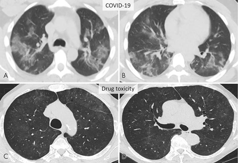
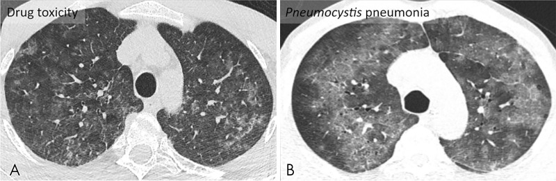
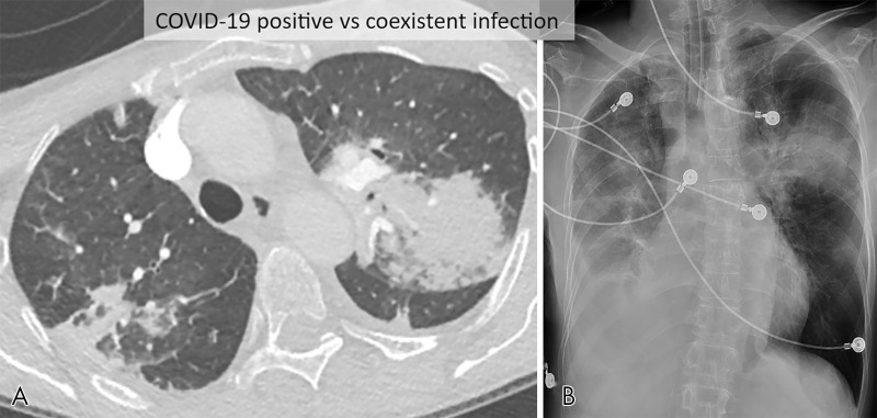
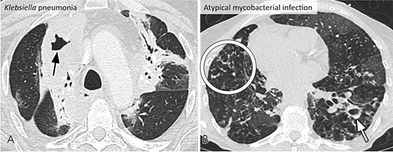
Routine screening CT for the identification of coronavirus disease 19 (COVID-19) pneumonia is currently not recommended by most radiology societies. However, the number of CT examinations performed in persons under investigation for COVID-19 has increased. We also anticipate that some patients will have incidentally detected findings that could be attributable to COVID-19 pneumonia, requiring radiologists to decide whether or not to mention COVID-19 specifically as a differential diagnostic possibility. We aim to provide guidance to radiologists in reporting CT findings potentially attributable to COVID-19 pneumonia, including standardized language to reduce reporting variability when addressing the possibility of COVID-19. When typical or indeterminate features of COVID-19 pneumonia are present in endemic areas as an incidental finding, we recommend contacting the referring providers to discuss the likelihood of viral infection. These incidental findings do not necessarily need to be reported as COVID-19 pneumonia. In this setting, using the term viral pneumonia can be a reasonable and inclusive alternative. However, if one opts to use the term COVID-19 in the incidental setting, consider the provided standardized reporting language. In addition, practice patterns may vary, and this document is meant to serve as a guide. Consultation with clinical colleagues at each institution is suggested to establish a consensus reporting approach. The goal of this expert consensus is to help radiologists recognize findings of COVID-19 pneumonia and aid their communication with other health care providers, assisting management of patients during this pandemic. Published under a CC BY 4.0 license.


Conflict of interest statement

Disclosures of Conflicts of Interest: S.S. Activities related to the present article: disclosed no relevant relationships. Activities not related to the present article: employed by University of Pennsylvania. Other relationships: disclosed no relevant relationships. F.U.K. Activities related to the present article: disclosed no relevant relationships. Activities not related to the present article: employed by University of Texas Southwestern Medical Center. Other relationships: disclosed no relevant relationships. S.A. Activities related to the present article: disclosed no relevant relationships. Activities not related to the present article: institution receives grants from NIH; author receives royalties from Elsevier for authoring of textbooks; Editor of RCTI (RSNA). Other relationships: disclosed no relevant relationships. S.B. disclosed no relevant relationships. J.H.C. disclosed no relevant relationships. M.C. disclosed no relevant relationships. T.S.H. disclosed no relevant relationships. J.P. Kanne Activities related to the present article: disclosed no relevant relationships. Activities not related to the present article: author is consultant for Parexel Informatics (clinical trial support, not related to topic at hand). Other relationships: disclosed no relevant relationships. S.K. disclosed no relevant relationships. J.P. Ko Activities related to the present article: disclosed no relevant relationships. Activities not related to the present article: institution receives money for consulting for Siemens Healthineers (research collaboration); author’s spouse is employed by Allovir (biotechnology company developing cell therapies for treatment of viral diseases). Other relationships: disclosed no relevant relationships. H.L. Activities related to the present article: disclosed no relevant relationships. Activities not related to the present article: institution receives grant from Siemens Healthineers; author receives travel/accommodations/meeting expenses from GE Healthcare and Edwards LifeSciences. Other relationships: editorial board member, RCTI.

Figures

Typical CT imaging features for COVID-19. Unenhanced thin-section axial images of the lungs in a 52-year-old man with a positive RT-PCR (A–D) show bilateral, multifocal rounded (asterisks) and peripheral GGO (arrows) with superimposed interlobular septal thickening and visible intralobular lines (“crazy-paving”). Routine screening CT for diagnosis or exclusion of COVID-19 is currently not recommended by most professional organizations or the U.S. Centers for Disease Control and Prevention.
Figure 1:
Typical CT imaging features for COVID-19. Unenhanced thin-section axial images of the lungs in a 52-year-old man with a positive RT-PCR (A–D) show bilateral, multifocal rounded (asterisks) and peripheral GGO (arrows) with superimposed interlobular septal thickening and visible intralobular lines (“crazy-paving”). Routine screening CT for diagnosis or exclusion of COVID-19 is currently not recommended by most professional organizations or the U.S. Centers for Disease Control and Prevention.
Typical CT imaging features for COVID-19. Unenhanced thin-section axial (A–C) and coronal multiplanar reformatted images (D) of the lungs in a 77-year-old man with a positive RT-PCR show bilateral, multifocal rounded and peripheral GGO. Routine screening CT for diagnosis or exclusion of COVID-19 is currently not recommended by most professional organizations or the U.S. Centers for Disease Control and Prevention.
Figure 2:
Typical CT imaging features for COVID-19. Unenhanced thin-section axial (A–C) and coronal multiplanar reformatted images (D) of the lungs in a 77-year-old man with a positive RT-PCR show bilateral, multifocal rounded and peripheral GGO. Routine screening CT for diagnosis or exclusion of COVID-19 is currently not recommended by most professional organizations or the U.S. Centers for Disease Control and Prevention.
Typical CT imaging features for COVID-19. Unenhanced axial (A–C) and sagittal multiplanar reformatted (D) images of the lungs in a 29-year-old man with a positive RT-PCR show multiple bilateral, rounded consolidations with surrounding GGO. Routine screening CT for diagnosis or exclusion of COVID-19 is currently not recommended by most professional organizations or the U.S. Centers for Disease Control and Prevention.
Figure 3:
Typical CT imaging features for COVID-19. Unenhanced axial (A–C) and sagittal multiplanar reformatted (D) images of the lungs in a 29-year-old man with a positive RT-PCR show multiple bilateral, rounded consolidations with surrounding GGO. Routine screening CT for diagnosis or exclusion of COVID-19 is currently not recommended by most professional organizations or the U.S. Centers for Disease Control and Prevention.
Typical CT imaging features for COVID-19 and other diseases with similar findings. Posterior, peripheral, and rounded GGO and consolidation in axial images of four patients; COVID-19 (A, B), organizing pneumonia secondary to dermatomyositis (C), and influenza A pneumonia (D). Organizing pneumonia and influenza pneumonia can be indistinguishable from COVID-19 by CT. Routine screening CT for diagnosis or exclusion of COVID-19 is currently not recommended by most professional organizations or the U.S. Centers for Disease Control and Prevention.
Figure 4:
Typical CT imaging features for COVID-19 and other diseases with similar findings. Posterior, peripheral, and rounded GGO and consolidation in axial images of four patients; COVID-19 (A, B), organizing pneumonia secondary to dermatomyositis (C), and influenza A pneumonia (D). Organizing pneumonia and influenza pneumonia can be indistinguishable from COVID-19 by CT. Routine screening CT for diagnosis or exclusion of COVID-19 is currently not recommended by most professional organizations or the U.S. Centers for Disease Control and Prevention.
Indeterminate CT imaging features for COVID-19. Unenhanced axial images in two patients showing patchy GGO with nonrounded morphology and no specific distribution, in a case of COVID-19 pneumonia (A, B) and acute lung injury from presumed drug toxicity (C, D). Routine screening CT for diagnosis or exclusion of COVID-19 is currently not recommended by most professional organizations or the U.S. Centers for Disease Control and Prevention.
Figure 5:
Indeterminate CT imaging features for COVID-19. Unenhanced axial images in two patients showing patchy GGO with nonrounded morphology and no specific distribution, in a case of COVID-19 pneumonia (A, B) and acute lung injury from presumed drug toxicity (C, D). Routine screening CT for diagnosis or exclusion of COVID-19 is currently not recommended by most professional organizations or the U.S. Centers for Disease Control and Prevention.
Indeterminate CT imaging features for COVID-19. Widespread GGO with nonrounded morphology and no specific distribution in unenhanced axial images from two different patients secondary to acute lung injury from presumed drug toxicity (A) and Pneumocystis pneumonia (B). Routine screening CT for diagnosis or exclusion of COVID-19 is currently not recommended by most professional organizations or the U.S. Centers for Disease Control and Prevention.
Figure 6:
Indeterminate CT imaging features for COVID-19. Widespread GGO with nonrounded morphology and no specific distribution in unenhanced axial images from two different patients secondary to acute lung injury from presumed drug toxicity (A) and Pneumocystis pneumonia (B). Routine screening CT for diagnosis or exclusion of COVID-19 is currently not recommended by most professional organizations or the U.S. Centers for Disease Control and Prevention.
Atypical CT imaging features for COVID-19. Contrast-enhanced axial CT image (A) and frontal chest radiograph (B) showing segmental consolidation without significant GGO. Although this patient tested positive for COVID-19, the imaging features are not typical and could represent pneumonia related to COVID-19 or a secondary infectious process. Routine screening CT for diagnosis or exclusion of COVID-19 is currently not recommended by most professional organizations or the U.S. Centers for Disease Control and Prevention.
Figure 7:
Atypical CT imaging features for COVID-19. Contrast-enhanced axial CT image (A) and frontal chest radiograph (B) showing segmental consolidation without significant GGO. Although this patient tested positive for COVID-19, the imaging features are not typical and could represent pneumonia related to COVID-19 or a secondary infectious process. Routine screening CT for diagnosis or exclusion of COVID-19 is currently not recommended by most professional organizations or the U.S. Centers for Disease Control and Prevention.
Atypical CT imaging features for COVID-19. Axial images of the lungs of two patients showing cavitation (arrow) in Klebsiella pneumonia (A) and tree-and-bud opacities (circle) and a cavity (arrow) in nontuberculous mycobacterial infection (B). Routine screening CT for diagnosis or exclusion of COVID-19 is currently not recommended by most professional organizations or the U.S. Centers for Disease Control and Prevention.
Figure 8:
Atypical CT imaging features for COVID-19. Axial images of the lungs of two patients showing cavitation (arrow) in Klebsiella pneumonia (A) and tree-and-bud opacities (circle) and a cavity (arrow) in nontuberculous mycobacterial infection (B). Routine screening CT for diagnosis or exclusion of COVID-19 is currently not recommended by most professional organizations or the U.S. Centers for Disease Control and Prevention.
Atypical CT imaging features for COVID-19. Axial CT images from two different patients showing tree-in-bud opacities and centrilobular nodules, caused by respiratory syncytial virus (A) and active tuberculosis (B). A small cavity (arrow) is also present in (B) Routine screening CT for diagnosis or exclusion of COVID-19 is currently not recommended by most professional organizations or the U.S. Centers for Disease Control and Prevention.
Figure 9:
Atypical CT imaging features for COVID-19. Axial CT images from two different patients showing tree-in-bud opacities and centrilobular nodules, caused by respiratory syncytial virus (A) and active tuberculosis (B). A small cavity (arrow) is also present in (B) Routine screening CT for diagnosis or exclusion of COVID-19 is currently not recommended by most professional organizations or the U.S. Centers for Disease Control and Prevention.

Similar articles

Cited by

References

    1. Naming the coronavirus disease (COVID-19) and the virus that causes it. https://www.who.int/emergencies/diseases/novel-coronavirus-2019/technica.... Published February 11, 2020. Accessed March 22, 2020.
    1. Gorbalenya AE, Baker SC, Baric RS, et al. Coronaviridae Study Group of the International Committee on Taxonomy of Viruses. The species Severe acute respiratory syndrome-related coronavirus: classifying 2019-nCoV and naming it SARS-CoV-2. Nat Microbiol 20205:536–544. - PMC - PubMed
    1. WHO Director-General’s opening remarks at the media briefing on COVID-19 - March 11, 2020. https://www.who.int/dg/speeches/detail/who-director-general-s-opening-re.... Published March 11, 2020. Accessed March 22, 2020.
    1. ACR Recommendations for the use of Chest Radiography and Computed Tomography (CT) for Suspected COVID-19 Infection. https://www.acr.org/Advocacy-and-Economics/ACR-Position-Statements/Recom.... Published March 11, 2020. Updated March 22, 2020. Accessed March 22, 2020.
    1. Society of Thoracic Radiology/American Society of Emergency Radiology COVID-19 Position Statement, March 11, 2020. https://thoracicrad.org. Published March 11, 2020. Accessed March 22, 2020.

LinkOut - more resources