

doi: 10.1148/ryct.2020200162.
eCollection 2020 Apr.
Coronavirus-HKU1 Pneumonia and Differential Diagnosis with COVID-19
Affiliations
- PMID: 33778574
- PMCID: PMC7233440
- DOI: 10.1148/ryct.2020200162
Item in Clipboard
Coronavirus-HKU1 Pneumonia and Differential Diagnosis with COVID-19
Radiol Cardiothorac Imaging.
.
No abstract available
Conflict of interest statement
Disclosures of Conflicts of Interest: E.P. disclosed no relevant relationships. F.D.S. disclosed no relevant relationships. M.C. disclosed no relevant relationships. A.P. disclosed no relevant relationships. N.F. disclosed no relevant relationships. F.A. disclosed no relevant relationships. D.L. disclosed no relevant relationships. S.C. disclosed no relevant relationships. P.C. disclosed no relevant relationships. V.S. disclosed no relevant relationships.
Figures
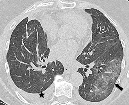
Images in 74-year-old woman with HKU1 coronavirus pneumonia. (a) CT scan performed at admission shows a large area of peripheral ground-glass opacity (arrow) in the lower left lobe and small pleural effusions (star). (b) The CT scan performed after 14 days shows almost complete resolution of the imaging findings.

Images in 74-year-old woman with HKU1 coronavirus pneumonia. (a) CT scan performed at admission shows a large area of peripheral ground-glass opacity (arrow) in the lower left lobe and small pleural effusions (star). (b) The CT scan performed after 14 days shows almost complete resolution of the imaging findings.

Images in patient with coronavirus disease 2019 (COVID-19) pneumonia. (a, b) Typical CT aspect of COVID-19 pneumonia: initial phase of disease with areas of ground-glass opacity peripherally (arrow in a); severe phase of disease with diffuse crazy paving pattern (arrow in b). Pleural effusions are absent.

Images in patient with coronavirus disease 2019 (COVID-19) pneumonia. (a, b) Typical CT aspect of COVID-19 pneumonia: initial phase of disease with areas of ground-glass opacity peripherally (arrow in a); severe phase of disease with diffuse crazy paving pattern (arrow in b). Pleural effusions are absent.
References
LinkOut - more resources
Full Text Sources
