. 2020 Jun 1;2(3):e200210.
doi: 10.1148/ryct.2020200210. eCollection 2020 Jun.

COVID-19: A Multimodality Review of Radiologic Techniques, Clinical Utility, and Imaging Features

Affiliations

COVID-19: A Multimodality Review of Radiologic Techniques, Clinical Utility, and Imaging Features

Sayan Manna et al. Radiol Cardiothorac Imaging. .

Abstract

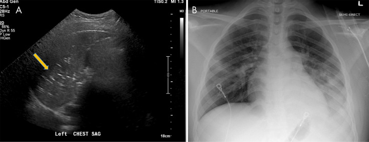
In this article we will review the imaging features of coronavirus disease 2019 (COVID-19) across multiple modalities, including radiography, CT, MRI, PET/CT, and US. Given that COVID-19 primarily affects the lung parenchyma by causing pneumonia, our directive is to focus on thoracic findings associated with COVID-19. We aim to enhance radiologists' understanding of this disease to help guide diagnosis and management. Supplemental material is available for this article. © RSNA, 2020.


Conflict of interest statement

Disclosures of Conflicts of Interest: S.M. disclosed no relevant relationships. J.W. Activities related to the present article: disclosed no relevant relationships. Activities not related to the present article: author is member of National Cancer Institute’s Physician Data Query board for cancer screening and prevention, unrelated to the topic of this article; participation is not reimbursed but travel to meetings is reimbursed. Other relationships: disclosed no relevant relationships. S.Z.M. disclosed no relevant relationships. D.T. disclosed no relevant relationships. N.V. disclosed no relevant relationships. M.F. disclosed no relevant relationships. M.A.C. disclosed no relevant relationships. J.D. disclosed no relevant relationships. C.E. disclosed no relevant relationships. A.J. disclosed no relevant relationships. M.C. disclosed no relevant relationships. A.B. disclosed no relevant relationships.

Figures

A, Posteroanterior chest radiograph in a 29-year-old woman with no past medical history, who presented to the emergency department with 4 days of fever, cough, pleuritic chest pain, diarrhea, and myalgias, shows faint peripheral rounded opacities in a lower lung distribution (arrows). B, Chest radiograph in a 40-year-old man with a history of diabetes mellitus who presented from a group home with fever, cough, and shortness of breath of duration 1 week demonstrates diffuse bilateral opacities with dense consolidation in the mid to lower lungs. C, Portable chest radiograph in an intubated 50-year-old man with a history of hypertension who presented with 1 week of fever, cough, congestion, and myalgias. On his 7th day of admission, 14 days after symptom onset, chest radiograph demonstrates multiple bilateral diffuse hazy pulmonary opacities. The patient was noted to have elevated inflammatory markers including C-reactive protein, interleukin-6, d-dimer, lactate dehydrogenase, and fibrinogen. All patients were confirmed positive for coronavirus disease 2019 with polymerase chain reaction testing, and the patient in C died two days later.
Figure 1:
A, Posteroanterior chest radiograph in a 29-year-old woman with no past medical history, who presented to the emergency department with 4 days of fever, cough, pleuritic chest pain, diarrhea, and myalgias, shows faint peripheral rounded opacities in a lower lung distribution (arrows). B, Chest radiograph in a 40-year-old man with a history of diabetes mellitus who presented from a group home with fever, cough, and shortness of breath of duration 1 week demonstrates diffuse bilateral opacities with dense consolidation in the mid to lower lungs. C, Portable chest radiograph in an intubated 50-year-old man with a history of hypertension who presented with 1 week of fever, cough, congestion, and myalgias. On his 7th day of admission, 14 days after symptom onset, chest radiograph demonstrates multiple bilateral diffuse hazy pulmonary opacities. The patient was noted to have elevated inflammatory markers including C-reactive protein, interleukin-6, d-dimer, lactate dehydrogenase, and fibrinogen. All patients were confirmed positive for coronavirus disease 2019 with polymerase chain reaction testing, and the patient in C died two days later.
Image in 42-year-old man with no past medical history other than obesity intubated in the intensive care unit. Chest radiograph demonstrates diffuse dense opacities with small bilateral pneumothoraces (arrows) in addition to severe bilateral neck and chest wall subcutaneous emphysema. The etiology of the air leak is unknown but suspected to be related to mechanical ventilation and high positive end-expiratory pressure settings, a finding that has been previously associated with acute respiratory distress syndrome. The patient was confirmed to be positive for coronavirus disease 2019 with polymerase chain reaction testing.
Figure 2:
Image in 42-year-old man with no past medical history other than obesity intubated in the intensive care unit. Chest radiograph demonstrates diffuse dense opacities with small bilateral pneumothoraces (arrows) in addition to severe bilateral neck and chest wall subcutaneous emphysema. The etiology of the air leak is unknown but suspected to be related to mechanical ventilation and high positive end-expiratory pressure settings, a finding that has been previously associated with acute respiratory distress syndrome. The patient was confirmed to be positive for coronavirus disease 2019 with polymerase chain reaction testing.
Corresponding axial chest CT scan without intravenous contrast material in patient in Figure 1 shows typical findings of coronavirus disease 2019 with peripheral and lower lobe–predominant dense consolidative opacities with a rounded morphology and mild surrounding ground-glass opacification (arrows).
Figure 3:
Corresponding axial chest CT scan without intravenous contrast material in patient in Figure 1 shows typical findings of coronavirus disease 2019 with peripheral and lower lobe–predominant dense consolidative opacities with a rounded morphology and mild surrounding ground-glass opacification (arrows).
A, Chest radiograph and B, axial non–contrast-enhanced chest CT scan in a 27-year-old man with no past medical history, who presented with a fever, vomiting, and myalgias of duration 3 days, demonstrate dense consolidation in the apicoposterior segment of the left upper lobe (arrows). The patient was confirmed to be positive for coronavirus disease 2019 with polymerase chain reaction testing. This single upper lobe involvement represents an atypical pattern of disease without any associated identifying factors in the patient’s history or presentation.
Figure 4:
A, Chest radiograph and B, axial non–contrast-enhanced chest CT scan in a 27-year-old man with no past medical history, who presented with a fever, vomiting, and myalgias of duration 3 days, demonstrate dense consolidation in the apicoposterior segment of the left upper lobe (arrows). The patient was confirmed to be positive for coronavirus disease 2019 with polymerase chain reaction testing. This single upper lobe involvement represents an atypical pattern of disease without any associated identifying factors in the patient’s history or presentation.
Axial chest CT scan following intravenous contrast material administration in the lower lungs of a 30-year-old male former smoker with a history of asthma who presented with fever, cough, and left lower back pain 3 weeks after symptom onset. CT demonstrates extensive dense patchy consolidation with relative subpleural sparing, most severe in the lung bases. Throughout the patient’s hospital course, he required supplemental oxygen via a nonrebreather mask and was noted to have elevated inflammatory markers, including C-reactive protein and interleukin-6. The patient was confirmed to be positive for coronavirus disease 2019 with polymerase chain reaction testing.
Figure 5:
Axial chest CT scan following intravenous contrast material administration in the lower lungs of a 30-year-old male former smoker with a history of asthma who presented with fever, cough, and left lower back pain 3 weeks after symptom onset. CT demonstrates extensive dense patchy consolidation with relative subpleural sparing, most severe in the lung bases. Throughout the patient’s hospital course, he required supplemental oxygen via a nonrebreather mask and was noted to have elevated inflammatory markers, including C-reactive protein and interleukin-6. The patient was confirmed to be positive for coronavirus disease 2019 with polymerase chain reaction testing.
Coronal chest CT scan in a 73-year-old woman with a history of hypertension and lupus and hospitalized for worsening respiratory status. Follow-up CT scan 30 days after initial symptoms demonstrates widespread reticulation with geographic regions of bronchiectasis and ground-glass opacity and clear demarcations between spared lobules, consistent with an organizing pattern.
Figure 6:
Coronal chest CT scan in a 73-year-old woman with a history of hypertension and lupus and hospitalized for worsening respiratory status. Follow-up CT scan 30 days after initial symptoms demonstrates widespread reticulation with geographic regions of bronchiectasis and ground-glass opacity and clear demarcations between spared lobules, consistent with an organizing pattern.
Image in 26-year-old woman with a recent history of sleeve gastrectomy complicated by splenic vein thrombosis who presented to the emergency department with left lower quadrant abdominal pain. T1-weighted axial MRI with intravenous contrast material of the abdomen and pelvis incidentally noted peripheral signal intensity abnormalities in the right lung base (arrow). The patient subsequently tested positive for coronavirus disease 2019 with polymerase chain reaction testing.
Figure 7:
Image in 26-year-old woman with a recent history of sleeve gastrectomy complicated by splenic vein thrombosis who presented to the emergency department with left lower quadrant abdominal pain. T1-weighted axial MRI with intravenous contrast material of the abdomen and pelvis incidentally noted peripheral signal intensity abnormalities in the right lung base (arrow). The patient subsequently tested positive for coronavirus disease 2019 with polymerase chain reaction testing.
Images in 33-year-old man with history of human immunodeficiency virus and treated Hodgkin lymphoma undergoing PET/CT for oncologic restaging. B, Fused PET/CT imaging shows bilateral peripheral-predominant intense sites of fluorodeoxyglucose avidity that correspond to ground-glass and mixed attenuation opacities on A, CT scans. A maximum standardized uptake value of 9.98 was seen. The patient was asymptomatic at time of imaging but presented to the emergency department 10 days later after developing hypoxia with 85% oxygen saturation on room air. The patient was confirmed to be positive for coronavirus disease 2019 with polymerase chain reaction testing.
Figure 8:
Images in 33-year-old man with history of human immunodeficiency virus and treated Hodgkin lymphoma undergoing PET/CT for oncologic restaging. B, Fused PET/CT imaging shows bilateral peripheral-predominant intense sites of fluorodeoxyglucose avidity that correspond to ground-glass and mixed attenuation opacities on A, CT scans. A maximum standardized uptake value of 9.98 was seen. The patient was asymptomatic at time of imaging but presented to the emergency department 10 days later after developing hypoxia with 85% oxygen saturation on room air. The patient was confirmed to be positive for coronavirus disease 2019 with polymerase chain reaction testing.
A, Lung US in a 17-year-old boy being treated for coronavirus disease 2019, on bilevel positive airway pressure in the pediatric intensive care unit, shows consolidation in the left lung with multiple echogenic foci representing “air bronchograms” (arrow). B, Chest radiograph in the same patient reveals multiple patchy consolidative pulmonary opacities most predominantly within the left lower lobe.
Figure 9:
A, Lung US in a 17-year-old boy being treated for coronavirus disease 2019, on bilevel positive airway pressure in the pediatric intensive care unit, shows consolidation in the left lung with multiple echogenic foci representing “air bronchograms” (arrow). B, Chest radiograph in the same patient reveals multiple patchy consolidative pulmonary opacities most predominantly within the left lower lobe.
Venous thromboembolic disease in coronavirus disease 2019 (COVID-19). A, Doppler US image of the left lower extremity in a 57-year-old man with COVID-19 demonstrates nonocclusive thrombus in the popliteal vein (arrow). B, Pelvic CT scan in a 33-year-old man demonstrates nonocclusive thrombus in the right common femoral vein (arrow). C, CT chest angiogram in a 69-year-old man demonstrates saddle pulmonary embolus (arrow).
Figure 10:
Venous thromboembolic disease in coronavirus disease 2019 (COVID-19). A, Doppler US image of the left lower extremity in a 57-year-old man with COVID-19 demonstrates nonocclusive thrombus in the popliteal vein (arrow). B, Pelvic CT scan in a 33-year-old man demonstrates nonocclusive thrombus in the right common femoral vein (arrow). C, CT chest angiogram in a 69-year-old man demonstrates saddle pulmonary embolus (arrow).
Arterial thrombosis in coronavirus disease 2019 (COVID-19). A, Sagittal CT angiogram of the abdomen in a 66-year-old woman with COVID-19 demonstrates thrombus in the celiac artery (arrow). B, Axial CT angiogram in the same patient demonstrates thrombus in both renal arteries (arrows) with associated hypoperfusion changes in both kidneys.
Figure 11:
Arterial thrombosis in coronavirus disease 2019 (COVID-19). A, Sagittal CT angiogram of the abdomen in a 66-year-old woman with COVID-19 demonstrates thrombus in the celiac artery (arrow). B, Axial CT angiogram in the same patient demonstrates thrombus in both renal arteries (arrows) with associated hypoperfusion changes in both kidneys.
Cerebral arterial thrombosis in coronavirus disease 2019 (COVID-19). A, Non–contrast-enhanced head CT scan in a 57-year-old man with COVID-19 demonstrates hypoattenuation throughout the left middle cerebral artery territory. B, CT angiogram in the same patient demonstrates focal occlusion of the left M2 branch (arrow) without distal reconstitution.
Figure 12:
Cerebral arterial thrombosis in coronavirus disease 2019 (COVID-19). A, Non–contrast-enhanced head CT scan in a 57-year-old man with COVID-19 demonstrates hypoattenuation throughout the left middle cerebral artery territory. B, CT angiogram in the same patient demonstrates focal occlusion of the left M2 branch (arrow) without distal reconstitution.

References

    1. Huang C, Wang Y, Li X, et al. . Clinical features of patients infected with 2019 novel coronavirus in Wuhan, China. Lancet 2020;395(10223):497–506 [Published correction appears in Lancet 2020;395(10223):496.]. - PMC - PubMed
    1. Coronavirus COVID-19 Global Cases by the Center for Systems Science and Engineering (CSSE) at Johns Hopkins University. (JHU). The Johns Hopkins University School of Medicine. https://coronavirus.jhu.edu/map.html. Published 2020. Accessed March 2020.
    1. Ai T, Yang Z, Hou H, et al. . Correlation of Chest CT and RT-PCR Testing in Coronavirus Disease 2019 (COVID-19) in China: A Report of 1014 Cases. Radiology 2020. 10.1148/radiol.2020200642. Published online February 26, 2020. - PMC - PubMed
    1. Rubin GD, Ryerson CJ, Haramati LB, et al. . The Role of Chest Imaging in Patient Management during the COVID-19 Pandemic: A Multinational Consensus Statement from the Fleischner Society. Radiology 2020. 10.1148/radiol.2020201365. Published online April 7, 2020. - PMC - PubMed
    1. Buonsenso D, Piano A, Raffaelli F, Bonadia N, de Gaetano Donati K, Franceschi F. Point-of-Care Lung Ultrasound findings in novel coronavirus disease-19 pnemoniae: a case report and potential applications during COVID-19 outbreak. Eur Rev Med Pharmacol Sci 2020;24(5):2776–2780. - PubMed

LinkOut - more resources