. 2020 Jul 9;2(4):e200289.
doi: 10.1148/ryct.2020200289. eCollection 2020 Aug.

Pulmonary Thromboembolism in COVID-19: Venous Thromboembolism or Arterial Thrombosis?

Affiliations

Pulmonary Thromboembolism in COVID-19: Venous Thromboembolism or Arterial Thrombosis?

Enrico Cavagna et al. Radiol Cardiothorac Imaging. .

Abstract

Purpose: To investigate CT pulmonary angiography findings of pulmonary thromboembolism (PTE) in coronavirus disease 2019 (COVID-19) and its association with clinical and radiologic conditions.

Materials and methods: This retrospective study includes 109 hospitalized patients with COVID-19 who underwent CT pulmonary angiography for suspected PTE from March 20 to May 3, 2020. Data were collected from our PACS. CT pulmonary angiography findings of PTE were evaluated. On the basis of the presence or absence of PTE, patients were divided into two groups, and their clinical and radiologic conditions were compared using the Mann-Whitney U test and χ2 test.

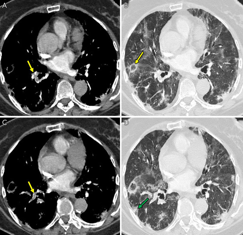
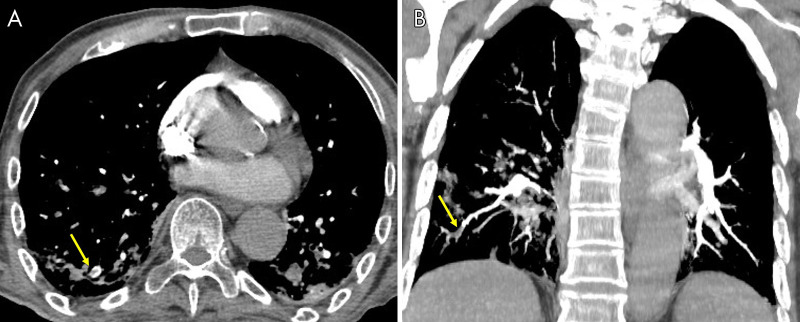
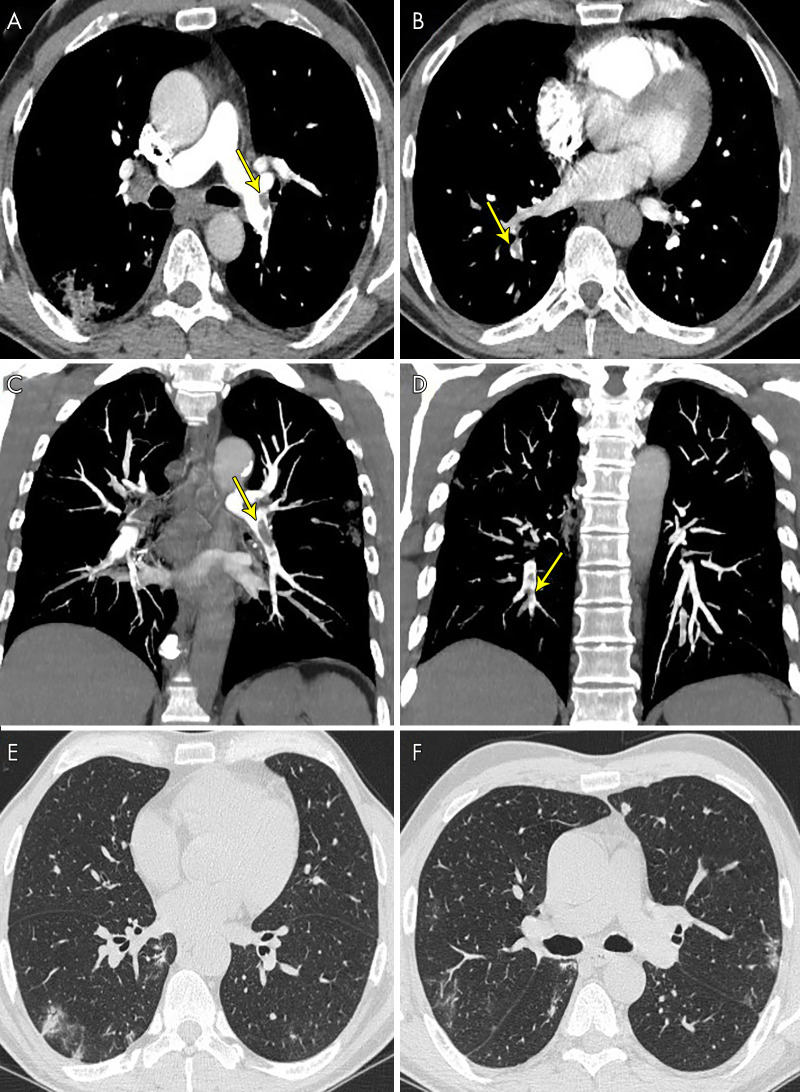
Results: The study population comprised 82 men and 19 women, with a mean age of 64.1 years ± 15.0 (95% confidence interval [CI]: 60.4, 67.6) years. CT pulmonary angiography was performed 19.8 days ± 6.1 (95% CI: 18.1, 20.2) after symptom onset and 10.5 days ± 3.8 (95% CI: 10.2, 12.9) after admission. Of 101 patients, 41 had PTE (40.6%). PTE was mostly bilateral or only right (37/41 [90.2%]), mainly involved segmental (37/41 [90.2%]) or subsegmental (25/41 [61.0%]) arteries and affected mainly the branches of the lower lobe (30/41 [73.2%]). Parenchymal segments supplied by segmental arteries with PTE showed a prevalent consolidation pattern (25/37 [67.6%]). Deep vein thrombosis was present only in five of 41 (12.2%) patients. Comparing groups with and without PTE, no significant difference was observed in age, sex, symptom onset, comorbidities, tumor history, use of respiratory supports, activated partial thromboplastin time, prothrombin time, and deep vein thrombosis. Conversely, differences were evaluated in CT lesion score (15.7 ± 1.4 [95% CI: 15.3, 16.1] vs 14.1 ± 1.1 [95% CI: 13.8, 14.4]; P = .035), d-dimer level (P < .001), lactate dehydrogenase level (P < .001), and C-reactive protein level (P = .042).

Conclusion: PTE in COVID-19 involves mainly the segmental and subsegmental arteries of segments affected by consolidations in patients with more severe lung disease. The authors hypothesize that the development of PTE in COVID-19 might be a pulmonary artery thrombosis because of severe lung inflammation and hypercoagulability rather than thromboembolism.© RSNA, 2020.


Conflict of interest statement

Disclosures of Conflicts of Interest: E.C. disclosed no relevant relationships. F.M. disclosed no relevant relationships. F.F. disclosed no relevant relationships.

Figures

An 85-year-old man, after 16 days of hospitalization in the intensive care unit with invasive ventilation, had a sudden increase in d-dimer value. The images show, A–C, pulmonary thromboembolism in the segmental arteries for the lower right lobe (yellow arrows). B, Lung parenchyma is characterized by reverse halo sign (yellow arrow) and, D, diffuse ground-glass and peripheral subpleural wedge-shaped consolidation in lower lung lobe (green arrow).
Figure 1:
An 85-year-old man, after 16 days of hospitalization in the intensive care unit with invasive ventilation, had a sudden increase in d-dimer value. The images show, A–C, pulmonary thromboembolism in the segmental arteries for the lower right lobe (yellow arrows). B, Lung parenchyma is characterized by reverse halo sign (yellow arrow) and, D, diffuse ground-glass and peripheral subpleural wedge-shaped consolidation in lower lung lobe (green arrow).
A 71-year-old woman, after 21 days of hospitalization with noninvasive ventilation, had sudden dyspnea and an increase in d-dimer value. The images show, A, pulmonary thromboembolism in the segmental and subsegmental arteries for the lower right lobe (yellow arrow), B, confirmed by a maximum intensity projection reconstruction (yellow arrow).
Figure 2:
A 71-year-old woman, after 21 days of hospitalization with noninvasive ventilation, had sudden dyspnea and an increase in d-dimer value. The images show, A, pulmonary thromboembolism in the segmental and subsegmental arteries for the lower right lobe (yellow arrow), B, confirmed by a maximum intensity projection reconstruction (yellow arrow).
A 63-year-old man, after 17 days of hospitalization without respiratory support, had a sudden increase in d-dimer value. The images show pulmonary thromboembolism, A, in the lobar arteries for the left lower lobe (yellow arrow) and, B, in the segmental arteries for the right lower lobe (yellow arrows), C, D, confirmed by maximum intensity projection reconstructions (yellow arrows). E, F, Lung parenchyma is characterized by some peripheral subpleural bilateral ground-glass opacities and consolidations.
Figure 3:
A 63-year-old man, after 17 days of hospitalization without respiratory support, had a sudden increase in d-dimer value. The images show pulmonary thromboembolism, A, in the lobar arteries for the left lower lobe (yellow arrow) and, B, in the segmental arteries for the right lower lobe (yellow arrows), C, D, confirmed by maximum intensity projection reconstructions (yellow arrows). E, F, Lung parenchyma is characterized by some peripheral subpleural bilateral ground-glass opacities and consolidations.

References

    1. Zhu N, Zhang D, Wang W, et al. . A novel coronavirus from patients with pneumonia in China, 2019. N Engl J Med 2020;382(8):727–733. - PMC - PubMed
    1. Ai T, Yang Z, Hou H, et al. . Correlation of chest CT and RT-PCR testing in coronavirus disease 2019 (COVID-19) in China: a report of 1014 cases. Radiology 2020. 10.1148/radiol.2020200642. Published online February 26, 2020. - PMC - PubMed
    1. Rubin GD, Ryerson CJ, Haramati LB, et al. . The role of chest imaging in patient management during the COVID-19 pandemic: a multinational consensus statement from the Fleischner Society. Radiology 2020;296(1):172–180. - PMC - PubMed
    1. Caruso D, Zerunian M, Polici M, et al. . Chest CT features of COVID-19 in Rome, Italy. Radiology 2020. 10.1148/radiol.2020201237. Published online April 3, 2020. - PMC - PubMed
    1. Terpos E, Ntanasis-Stathopoulos I, Elalamy I, et al. . Hematological findings and complications of COVID-19. Am J Hematol 2020;95(7):834–847. - PMC - PubMed