. 2020 Jul 23;2(4):e200312.
doi: 10.1148/ryct.2020200312. eCollection 2020 Aug.

Diagnostic Accuracy of North America Expert Consensus Statement on Reporting CT Findings in Patients Suspected of Having COVID-19 Infection: An Italian Single-Center Experience

Affiliations

Diagnostic Accuracy of North America Expert Consensus Statement on Reporting CT Findings in Patients Suspected of Having COVID-19 Infection: An Italian Single-Center Experience

Federica Ciccarese et al. Radiol Cardiothorac Imaging. .

Abstract

Purpose: To evaluate the diagnostic accuracy of the four standardized categories for CT reporting proposed by the Radiological Society of North America (RSNA) to support a faster triage compared with real-time reverse-transcription polymerase chain reaction (RT-PCR), which is the reference standard for suspected coronavirus disease 2019 (COVID-19), but has long reporting time (6-48 hours).

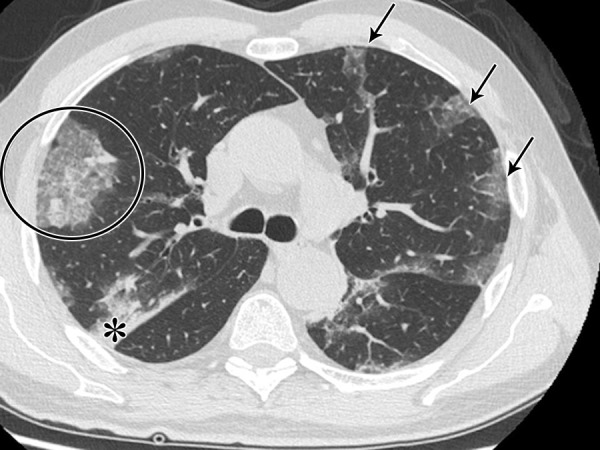
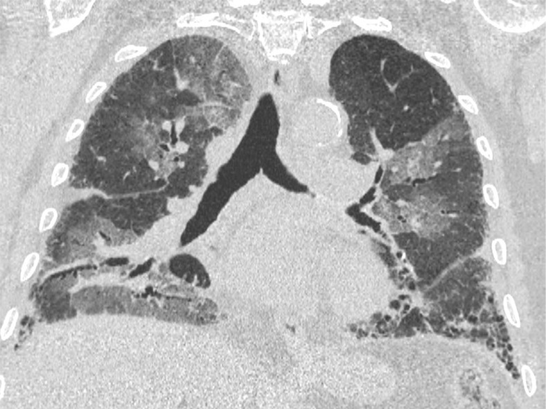
Materials and methods: A retrospective analysis of 569 thin-section CT examinations performed for patients suspected of having COVID-19 from February 27 to March 27, 2020 (peak of infection in Italy) was conducted. The imaging pattern was classified according to the statement by the RSNA as "typical," "indeterminate," "atypical," and "negative" and compared with RT-PCR for 460 patients. Interobserver variability in reporting between a senior and a junior radiologist was evaluated. Use of the vascular enlargement sign in indeterminate cases was also assessed.

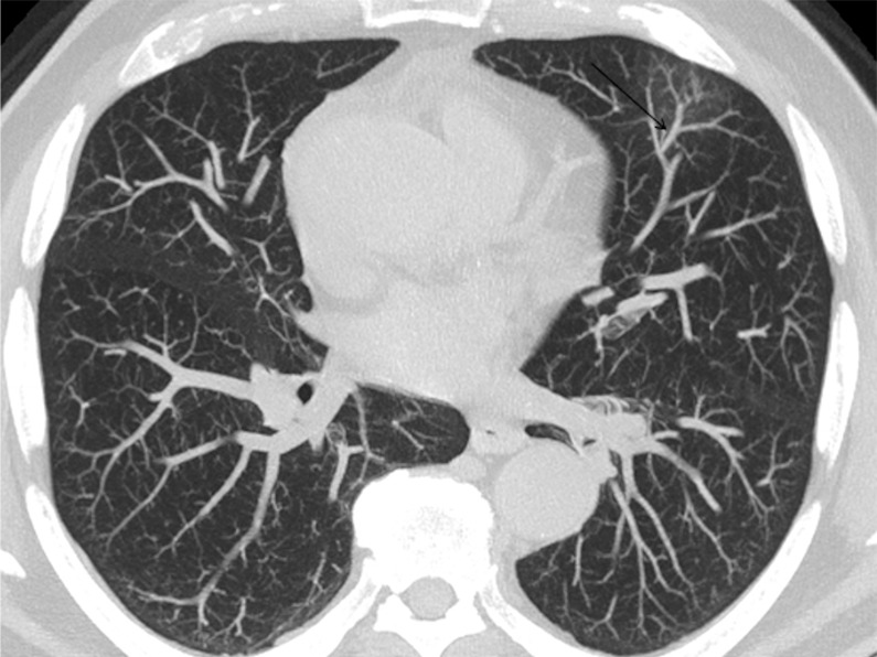
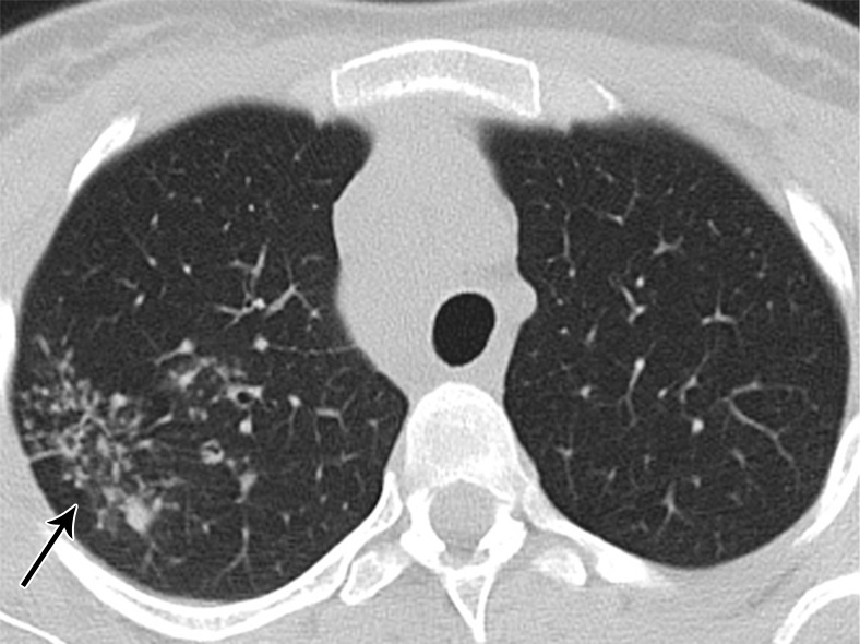
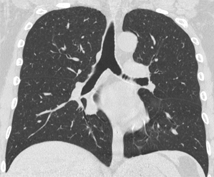
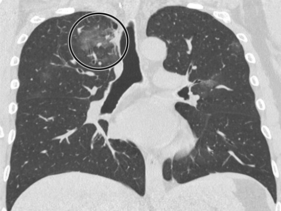
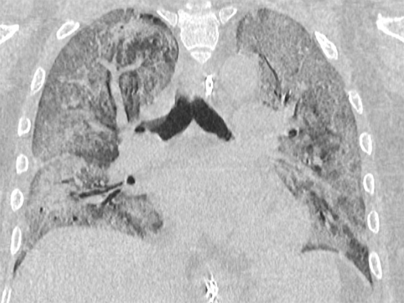
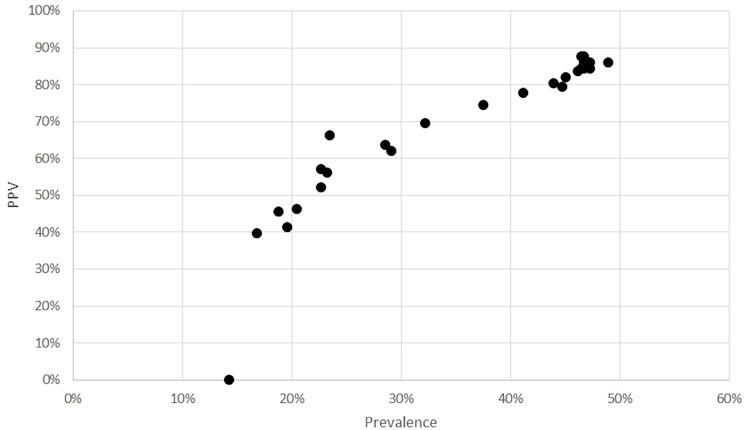
Results: The diagnosis of COVID-19 was made in 45.9% (211/460) of patients. The "typical" pattern (n = 172) showed a sensitivity of 71.6%, a specificity of 91.6%, and a positive predictive value of 87.8% for COVID-19. The "atypical" (n = 67) and "negative" (n = 123) pattern demonstrated a positive predictive value of 89.6% and 86.2% for non-COVID-19, respectively. The "indeterminate" (n = 98) pattern was nonspecific, but vascular enlargement was most frequently found in patients with COVID-19 (86.1%; P < .001). Interobserver agreement was good for the "typical" and "negative" pattern and fair for "indeterminate" and "atypical" (κ = 0.5; P = .002).

Conclusion: In an epidemic setting, the application of the four categories proposed by the RSNA provides a standardized diagnostic hypothesis, strongly linked to the RT-PCR results for the "typical," "atypical," and "negative" pattern. In the "indeterminate" pattern, the analysis of the vascular enlargement sign could facilitate the interpretation of imaging features.© RSNA, 2020.


Conflict of interest statement

Disclosures of Conflicts of Interest: F. Ciccarese disclosed no relevant relationships. F. Coppola disclosed no relevant relationships. D.S. disclosed no relevant relationships. G.L.G. disclosed no relevant relationships. V.L. disclosed no relevant relationships. A.P. disclosed no relevant relationships. C.D.B. disclosed no relevant relationships. C.B. disclosed no relevant relationships. R.G. disclosed no relevant relationships.

Figures

Flowchart of the study.
Figure 1:
Flowchart of the study.
The “typical” pattern in a 62-year-old man with COVID-19, characterized by bilateral ground-glass opacities peripheral in distribution (arrows), consolidations (*), and crazy paving (circle).
Figure 2:
The “typical” pattern in a 62-year-old man with COVID-19, characterized by bilateral ground-glass opacities peripheral in distribution (arrows), consolidations (*), and crazy paving (circle).
A 74-year-old man arriving at the emergency department with fever. (a) The CT scan documented a focal ground-glass opacity in the superior segment of the lingula (circle), classified as an “indeterminate” pattern. Notably, there was a vascular enlargement in the pulmonary artery branch afferent to the lesion, as (b) the maximum intensity projection better demonstrated (arrow). The real-time reverse-transcription polymerase chain reaction test was positive for severe acute respiratory syndrome coronavirus 2 infection.
Figure 3a:
A 74-year-old man arriving at the emergency department with fever. (a) The CT scan documented a focal ground-glass opacity in the superior segment of the lingula (circle), classified as an “indeterminate” pattern. Notably, there was a vascular enlargement in the pulmonary artery branch afferent to the lesion, as (b) the maximum intensity projection better demonstrated (arrow). The real-time reverse-transcription polymerase chain reaction test was positive for severe acute respiratory syndrome coronavirus 2 infection.
Figure 3b:
A 74-year-old man arriving at the emergency department with fever. (a) The CT scan documented a focal ground-glass opacity in the superior segment of the lingula (circle), classified as an “indeterminate” pattern. Notably, there was a vascular enlargement in the pulmonary artery branch afferent to the lesion, as (b) the maximum intensity projection better demonstrated (arrow). The real-time reverse-transcription polymerase chain reaction test was positive for severe acute respiratory syndrome coronavirus 2 infection.
An 89-year-old man with a known interstitial lung disease, arriving at the emergency department with dyspnea. (a) The CT scan documented a diffuse ground-glass opacity, without a specific distribution, an “indeterminate” pattern. The real-time reverse-transcription polymerase chain reaction test was positive for severe acute respiratory syndrome coronavirus 2. In addition, in this case, (b) dilatation of pulmonary vessels was seen (arrow).
Figure 4a:
An 89-year-old man with a known interstitial lung disease, arriving at the emergency department with dyspnea. (a) The CT scan documented a diffuse ground-glass opacity, without a specific distribution, an “indeterminate” pattern. The real-time reverse-transcription polymerase chain reaction test was positive for severe acute respiratory syndrome coronavirus 2. In addition, in this case, (b) dilatation of pulmonary vessels was seen (arrow).
Figure 4b:
An 89-year-old man with a known interstitial lung disease, arriving at the emergency department with dyspnea. (a) The CT scan documented a diffuse ground-glass opacity, without a specific distribution, an “indeterminate” pattern. The real-time reverse-transcription polymerase chain reaction test was positive for severe acute respiratory syndrome coronavirus 2. In addition, in this case, (b) dilatation of pulmonary vessels was seen (arrow).
A 26-year-old woman arriving at the emergency department with persistent cough. (a) The CT scan demonstrated an “atypical” pattern characterized by small peribronchial consolidations with a tree-in-bud appearance located in the apical segment of the superior right lobe (arrow) and (b) multiple right hilar and mediastinal lymphadenopathy (circle). The final diagnosis was of lung and nodal tuberculosis.
Figure 5a:
A 26-year-old woman arriving at the emergency department with persistent cough. (a) The CT scan demonstrated an “atypical” pattern characterized by small peribronchial consolidations with a tree-in-bud appearance located in the apical segment of the superior right lobe (arrow) and (b) multiple right hilar and mediastinal lymphadenopathy (circle). The final diagnosis was of lung and nodal tuberculosis.
Figure 5b:
A 26-year-old woman arriving at the emergency department with persistent cough. (a) The CT scan demonstrated an “atypical” pattern characterized by small peribronchial consolidations with a tree-in-bud appearance located in the apical segment of the superior right lobe (arrow) and (b) multiple right hilar and mediastinal lymphadenopathy (circle). The final diagnosis was of lung and nodal tuberculosis.
A 47-year-old man arriving at the emergency department with fever and diarrhea. (a) The baseline CT scan did not show findings suggestive for a pneumonia-“negative” pattern, but the real-time polymerase chain reaction test was positive for severe acute respiratory syndrome coronavirus 2 infection. (b) After 5 days, owing to a worsening of the clinical conditions, CT was repeated, revealing a “typical” pattern characterized by bilateral ground-glass opacities (circle). (c) After 12 days (17 days from the onset of symptoms), further radiologic progression with development of diffuse alveolar damage and acute respiratory distress syndrome would lead to the death of the patient.
Figure 6a:
A 47-year-old man arriving at the emergency department with fever and diarrhea. (a) The baseline CT scan did not show findings suggestive for a pneumonia-“negative” pattern, but the real-time polymerase chain reaction test was positive for severe acute respiratory syndrome coronavirus 2 infection. (b) After 5 days, owing to a worsening of the clinical conditions, CT was repeated, revealing a “typical” pattern characterized by bilateral ground-glass opacities (circle). (c) After 12 days (17 days from the onset of symptoms), further radiologic progression with development of diffuse alveolar damage and acute respiratory distress syndrome would lead to the death of the patient.
Figure 6b:
A 47-year-old man arriving at the emergency department with fever and diarrhea. (a) The baseline CT scan did not show findings suggestive for a pneumonia-“negative” pattern, but the real-time polymerase chain reaction test was positive for severe acute respiratory syndrome coronavirus 2 infection. (b) After 5 days, owing to a worsening of the clinical conditions, CT was repeated, revealing a “typical” pattern characterized by bilateral ground-glass opacities (circle). (c) After 12 days (17 days from the onset of symptoms), further radiologic progression with development of diffuse alveolar damage and acute respiratory distress syndrome would lead to the death of the patient.
Figure 6c:
A 47-year-old man arriving at the emergency department with fever and diarrhea. (a) The baseline CT scan did not show findings suggestive for a pneumonia-“negative” pattern, but the real-time polymerase chain reaction test was positive for severe acute respiratory syndrome coronavirus 2 infection. (b) After 5 days, owing to a worsening of the clinical conditions, CT was repeated, revealing a “typical” pattern characterized by bilateral ground-glass opacities (circle). (c) After 12 days (17 days from the onset of symptoms), further radiologic progression with development of diffuse alveolar damage and acute respiratory distress syndrome would lead to the death of the patient.
Positive predictive value for a “typical” pattern in relation with prevalence.
Figure 7:
Positive predictive value for a “typical” pattern in relation with prevalence.

References

    1. WHO Coronavirus disease (COVID-2019) situation reports-147 . World Health Organization website. https://www.who.int/emergencies/diseases/novel-coronavirus-2019/situatio.... Published June 15, 2020.
    1. Rubin GD, Ryerson CJ, Haramati LB, et al. The Role of Chest Imaging in Patient Management During the COVID-19 Pandemic: A Multinational Consensus Statement From the Fleischner Society. Chest 2020;158(1):106–116. - PMC - PubMed
    1. Ai T, Yang Z, Hou H, et al. Correlation of Chest CT and RT-PCR Testing in Coronavirus Disease 2019 (COVID-19) in China: A Report of 1014 Cases. Radiology 2020;296(2):E32–E40. - PMC - PubMed
    1. Li Y, Yao L, Li J, et al. Stability issues of RT-PCR testing of SARS-CoV-2 for hospitalized patients clinically diagnosed with COVID-19. J Med Virol 2020;92(7):903–908. - PMC - PubMed
    1. Wong HYF, Lam HYS, Fong AH, et al. Frequency and Distribution of Chest Radiographic Findings in COVID-19 Positive Patients. Radiology 2020;296(2):E72–E78. - PMC - PubMed

LinkOut - more resources