

A Characteristic Chest Radiographic Pattern in the Setting of the COVID-19 Pandemic
- PMID: 33778626
- PMCID: PMC7605076
- DOI: 10.1148/ryct.2020200280
A Characteristic Chest Radiographic Pattern in the Setting of the COVID-19 Pandemic
Abstract
Purpose: To determine the utility of chest radiography in aiding clinical diagnosis of coronavirus disease 2019 (COVID-19) utilizing reverse-transcription polymerase chain reaction (RT-PCR) as the standard of comparison.
Materials and methods: A retrospective study was performed of persons under investigation for COVID-19 presenting to this institution during the exponential growth phase of the COVID-19 outbreak in New Orleans (March 13-25, 2020). Three hundred seventy-six in-hospital chest radiographic examinations for 366 individual patients were reviewed along with concurrent RT-PCR tests. Two experienced radiologists categorized each chest radiograph as characteristic, nonspecific, or negative in appearance for COVID-19, utilizing well-documented COVID-19 imaging patterns. Chest radiograph categorization was compared against RT-PCR results to determine the utility of chest radiography in diagnosing COVID-19.
Results: Of the 366 patients, the study consisted of 178 male (49%) and 188 female (51%) patients with a mean age of 52.7 years (range, 17 to 98 years). Of the 376 chest radiographic examinations, 37 (10%) exhibited the characteristic COVID-19 appearance; 215 (57%) exhibited the nonspecific appearance; and 124 (33%) were considered negative for a pulmonary abnormality. Of the 376 RT-PCR tests evaluated, 200 (53%) were positive and 176 (47%) were negative. RT-PCR tests took an average of 2.5 days ± 0.7 to provide results. Sensitivity and specificity for correctly identifying COVID-19 with a characteristic chest radiographic pattern was 15.5% (31/200) and 96.6% (170/176), with a positive predictive value and negative predictive value of 83.8% (31/37) and 50.1% (170/339), respectively.
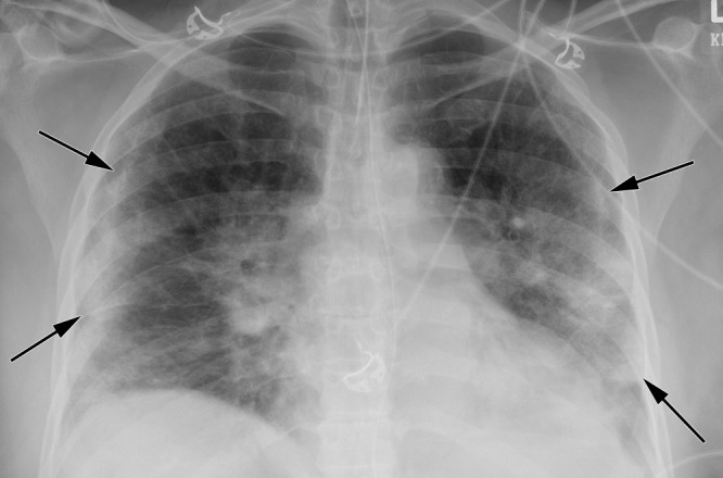
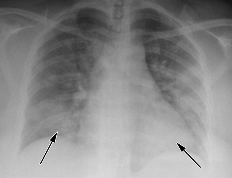
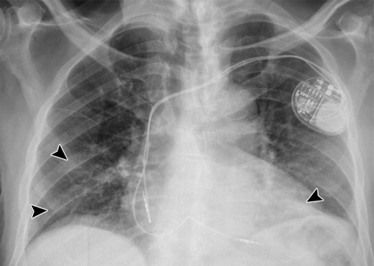
Conclusion: The presence of patchy and/or confluent, bandlike ground-glass opacity or consolidation in a peripheral and mid to lower lung zone distribution on a chest radiograph obtained in the setting of pandemic COVID-19 was highly suggestive of severe acute respiratory syndrome coronavirus 2 infection and should be used in conjunction with clinical judgment to make a diagnosis.© RSNA, 2020.
2021 by the Radiological Society of North America, Inc.
Conflict of interest statement
Disclosures of Conflicts of Interest: D.L.S. Activities related to the present article: disclosed no relevant relationships. Activities not related to the present article: author provides expert testimony for Hall Booth Smith on a matter unrelated to this article; author is paid by Genentech for speakers bureau giving nonbranded lectures on interstitial lung disease (unrelated to this article). Other relationships: disclosed no relevant relationships. J.P.G. disclosed no relevant relationships. C.B. disclosed no relevant relationships. B.S. disclosed no relevant relationships.
Figures


References
-
- COVID-19 Dashboard by the Center for Systems Science and Engineering (CSSE). https://coronavirus.jhu.edu/map.html. Accessed April 21, 2020.
-
- Zhou S, Wang Y, Zhu T, Xia L. CT Features of Coronavirus Disease 2019 (COVID-19) Pneumonia in 62 Patients in Wuhan, China. AJR Am J Roentgenol 2020;214(6):1287–1294. - PubMed
-
- Cheng Z, Lu Y, Cao Q, et al. Clinical Features and Chest CT Manifestations of Coronavirus Disease 2019 (COVID-19) in a Single-Center Study in Shanghai, China. AJR Am J Roentgenol 2020;215(1):121–126. - PubMed
-
- Simpson S, Kay FU, Abbara S, et al. Radiological Society of North America Expert Consensus Statement on Reporting Chest CT Findings Related to COVID-19. Endorsed by the Society of Thoracic Radiology, the American College of Radiology, and RSNA - Secondary Publication. J Thorac Imaging 2020;35(4):219–227. - PMC - PubMed
LinkOut - more resources
Full Text Sources
