Case Reports
. 2020 Dec 10;2(6):e200417.
doi: 10.1148/ryct.2020200417. eCollection 2020 Dec.

Esophagopericardial Fistula Causing Pyopneumopericardium

Affiliations
Case Reports

Esophagopericardial Fistula Causing Pyopneumopericardium

Andrew W Bowman et al. Radiol Cardiothorac Imaging. .

Abstract

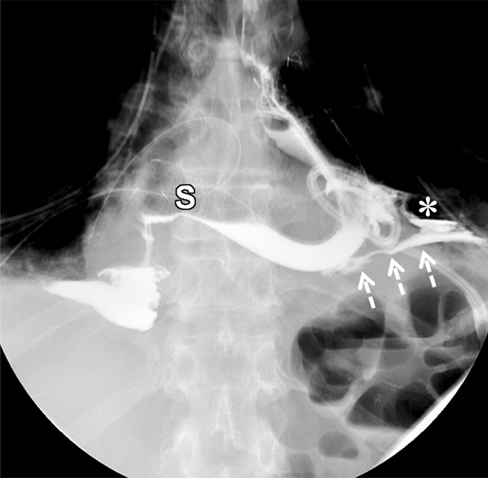
An esophagopericardial fistula is a rare complication of esophageal malignancy, trauma, or surgery. Imaging is a cornerstone of diagnosis, with detection of pneumopericardium or hydropneumopericardium at imaging raising suspicion for pyopneumopericardium and prompting immediate search for the causative pathologic process. Given the high associated mortality rate of over 50% for patients with esophagopericardial fistulas, early diagnosis and intervention are vital. Supplemental material is available for this article. © RSNA, 2020.


Conflict of interest statement

Disclosures of Conflicts of Interest: A.W.B. disclosed no relevant relationships. D.J.D. disclosed no relevant relationships. R.T.F. disclosed no relevant relationships.

Figures

(a) Frontal and (b) lateral radiographs demonstrate widening of the cardiac silhouette. Note air on both sides outlining the pericardium on the frontal view (thin arrows), and an air-fluid layer in the anterior chest on lateral view (thick arrows), indicative of hydropneumopericardium. Pleural effusions and a left lower thorax surgical drain are also present.
Figure 1a:
(a) Frontal and (b) lateral radiographs demonstrate widening of the cardiac silhouette. Note air on both sides outlining the pericardium on the frontal view (thin arrows), and an air-fluid layer in the anterior chest on lateral view (thick arrows), indicative of hydropneumopericardium. Pleural effusions and a left lower thorax surgical drain are also present.
Figure 1b:
(a) Frontal and (b) lateral radiographs demonstrate widening of the cardiac silhouette. Note air on both sides outlining the pericardium on the frontal view (thin arrows), and an air-fluid layer in the anterior chest on lateral view (thick arrows), indicative of hydropneumopericardium. Pleural effusions and a left lower thorax surgical drain are also present.
Axial CT image of the chest without intravenous contrast but with oral contrast demonstrates an air-fluid level (thick arrows) within the pericardial sac (thin arrows), confirming the large hydropneumopericardium. Oral contrast is seen in the lumen of the visualized alimentary tract (L) as well as a loculated extraluminal collection (*), consistent with a chronic anastomotic leak. Bilateral small pleural effusions are also present, left larger than right.
Figure 2:
Axial CT image of the chest without intravenous contrast but with oral contrast demonstrates an air-fluid level (thick arrows) within the pericardial sac (thin arrows), confirming the large hydropneumopericardium. Oral contrast is seen in the lumen of the visualized alimentary tract (L) as well as a loculated extraluminal collection (*), consistent with a chronic anastomotic leak. Bilateral small pleural effusions are also present, left larger than right.
Frontal images from an esophagogram performed with water-soluble contrast material after surgical drainage of the pericardium and subsequent placement of an esophageal stent (S) across the esophagojejunostomy. (a) Early image in the study demonstrates persistent extraluminal contrast (dashed arrows) outside the alimentary tract, coursing toward midline. The loculated extraluminal collection depicted in Figure 2 remains and is again seen (*). (b) More delayed image from the study demonstrates oral contrast filling the pericardial space (thin arrows), confirming the presence of a persistent esophagopericardial fistula. Bilateral surgical drains are also present.
Figure 3a:
Frontal images from an esophagogram performed with water-soluble contrast material after surgical drainage of the pericardium and subsequent placement of an esophageal stent (S) across the esophagojejunostomy. (a) Early image in the study demonstrates persistent extraluminal contrast (dashed arrows) outside the alimentary tract, coursing toward midline. The loculated extraluminal collection depicted in Figure 2 remains and is again seen (*). (b) More delayed image from the study demonstrates oral contrast filling the pericardial space (thin arrows), confirming the presence of a persistent esophagopericardial fistula. Bilateral surgical drains are also present.
Figure 3b:
Frontal images from an esophagogram performed with water-soluble contrast material after surgical drainage of the pericardium and subsequent placement of an esophageal stent (S) across the esophagojejunostomy. (a) Early image in the study demonstrates persistent extraluminal contrast (dashed arrows) outside the alimentary tract, coursing toward midline. The loculated extraluminal collection depicted in Figure 2 remains and is again seen (*). (b) More delayed image from the study demonstrates oral contrast filling the pericardial space (thin arrows), confirming the presence of a persistent esophagopericardial fistula. Bilateral surgical drains are also present.

References

    1. Ladurner R, Qvick LM, Hohenbleicher F, Hallfeldt KK, Mutschler W, Mussack T. Pneumopericardium in blunt chest trauma after high-speed motor vehicle accidents. Am J Emerg Med 2005;23(1):83–86. - PubMed
    1. Farkas ZC, Pal S, Jolly GP, Lim MMD, Malik A, Malekan R. Esophagopericardial fistula and pneumopericardium from caustic ingestion and esophageal stent. Ann Thorac Surg 2019;107(3):e207–e208. - PubMed
    1. Dagres N, Kottkamp H, Piorkowski C, et al. Rapid detection and successful treatment of esophageal perforation after radiofrequency ablation of atrial fibrillation: lessons from five cases. J Cardiovasc Electrophysiol 2006;17(11):1213–1215. - PubMed
    1. Bunch TJ, Nelson J, Foley T, et al. Temporary esophageal stenting allows healing of esophageal perforations following atrial fibrillation ablation procedures. J Cardiovasc Electrophysiol 2006;17(4):435–439. - PubMed
    1. Möschler O, Nies C, Mueller MK. Endoscopic vacuum therapy for esophageal perforations and leakages. Endosc Int Open 2015;3(6):E554–E558. - PMC - PubMed

Publication types

LinkOut - more resources