Review
. 2021 Feb 11;3(1):e200314.
doi: 10.1148/ryct.2021200314. eCollection 2021 Feb.

Chronic Lung Allograft Dysfunction: Review of CT and Pathologic Findings

Affiliations
Review

Chronic Lung Allograft Dysfunction: Review of CT and Pathologic Findings

Danielle Byrne et al. Radiol Cardiothorac Imaging. .

Abstract

Chronic lung allograft dysfunction (CLAD) is the most common cause of mortality in lung transplant recipients after the 1st year of transplantation. CLAD has traditionally been classified into two distinct obstructive and restrictive forms: bronchiolitis obliterans syndrome and restrictive allograft syndrome. However, CLAD may manifest with a spectrum of imaging and pathologic findings and a combination of obstructive and restrictive physiologic abnormalities. Although the initial CT manifestations of CLAD may be nonspecific, the progression of findings at follow-up should signal the possibility of CLAD and may be present on imaging studies prior to the development of functional abnormalities of the lung allograft. This review encompasses the evolution of CT findings in CLAD, with emphasis on the underlying pathogenesis and pathologic condition, to enhance understanding of imaging findings. The purpose of this article is to familiarize the radiologist with the initial and follow-up CT findings of the obstructive, restrictive, and mixed forms of CLAD, for which early diagnosis and treatment may result in improved survival. Supplemental material is available for this article. © RSNA, 2021.


Conflict of interest statement

Disclosures of Conflicts of Interest: D.B. disclosed no relevant relationships. R.G.N. disclosed no relevant relationships. J.C.E. disclosed no relevant relationships. J.Y. disclosed no relevant relationships. R.L. disclosed no relevant relationships. C.B. disclosed no relevant relationships. J.R.S. disclosed no relevant relationships. O.M.M. disclosed no relevant relationships. N.L.M. disclosed no relevant relationships. A.M.B. disclosed no relevant relationships.

Figures

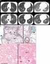
Obstructive chronic lung allograft dysfunction in a 61-year-old man. A, No airtrapping on CT images obtained on posttransplant day 221, with appropriate increased bilateral attenuation on expiratory (EXP) images evolving to, B, severe airtrapping on CT images obtained on posttransplant day 991, manifested by bilateral areas of increasing lucency at expiration. C, At transbronchial biopsy specimen assessment, subepithelial connective tissue (arrows) were apparent in portions of these two membranous bronchioles, identifiable because of their smooth muscle walls (arrowheads on C). Although hemosiderin-containing macrophages can be seen in some airspaces in the lower portion of the biopsy specimen, the alveolar walls are of relatively normal thickness. Bar = 0.5 mm. Additional patient information is found in Tables E1 and E2 (supplement). INS = inspiratory.
Figure 1:
Obstructive chronic lung allograft dysfunction in a 61-year-old man. A, No airtrapping on CT images obtained on posttransplant day 221, with appropriate increased bilateral attenuation on expiratory (EXP) images evolving to, B, severe airtrapping on CT images obtained on posttransplant day 991, manifested by bilateral areas of increasing lucency at expiration. C, At transbronchial biopsy specimen assessment, subepithelial connective tissue (arrows) were apparent in portions of these two membranous bronchioles, identifiable because of their smooth muscle walls (arrowheads on C). Although hemosiderin-containing macrophages can be seen in some airspaces in the lower portion of the biopsy specimen, the alveolar walls are of relatively normal thickness. Bar = 0.5 mm. Additional patient information is found in Tables E1 and E2 (supplement). INS = inspiratory.
A, Mixed-phenotype chronic lung allograft dysfunction in a 30-year-old man. CT images obtained on posttransplant day 1833 show predominantly peribronchovascular reticulation with nodular thickening of the interlobular septa (ILS), perilymphatic nodules, and associated architectural distortion. The expiratory image obtained on day 1833 demonstrates new moderate airtrapping with a lower-than-expected increase in attenuation and lack of volume reduction. B, Low-power images of the left lower lobe from the explant in A. The pleura (P) shows mild fibrosis. Bands of fibrous tissue, presumably the residua of unresolved organizing pneumonia, extend from the subpleural zone and penetrate the underlying parenchyma following the ILS (arrows). Some of the centrilobular airways demonstrate changes of constrictive obliterative bronchiolitis (arrowheads). Movat pentachrome staining is used. The horizontal bar is equivalent to 2 mm. C, From a different area of the left lower lobe, this medium-power micrograph of a membranous bronchiole (Br) demonstrates the classic finding of constrictive obliterative bronchiolitis, in which the subepithelial zone, of normal to negligible thickness, is expanded by a myofibroblastic matrix (double arrow), causing a severe reduction in luminal patency. Movat pentachrome staining is used. The horizontal bar is equivalent to 0.5 mm. D, Microscopic section from a chronic lung allograft dysfunction explant shows uniform alveolar wall thickening and architectural sparing changes of nonspecific interstitial pneumonia. The airspaces contain scattered inflammatory cells and occasionally contain some edematous fluid. The horizontal bar is equivalent to 0.5 mm. E, Superimposed on a background of nonspecific interstitial pneumonia, the parenchyma shows a combination of alveolar macrophage accumulation, simulating a desquamative interstitial pneumonia pattern (*) with airspace cholesterol granulomas (arrow), suggesting a component of obstructive pneumonitis. Some of the tissue compaction is likely due to atelectasis. Hematoxylin-eosin staining is used. Additional patient information is found in Tables E1 and E2 (supplement). ils = interlobular septal thickening, PA = pulmonary artery.
Figure 2:
A, Mixed-phenotype chronic lung allograft dysfunction in a 30-year-old man. CT images obtained on posttransplant day 1833 show predominantly peribronchovascular reticulation with nodular thickening of the interlobular septa (ILS), perilymphatic nodules, and associated architectural distortion. The expiratory image obtained on day 1833 demonstrates new moderate airtrapping with a lower-than-expected increase in attenuation and lack of volume reduction. B, Low-power images of the left lower lobe from the explant in A. The pleura (P) shows mild fibrosis. Bands of fibrous tissue, presumably the residua of unresolved organizing pneumonia, extend from the subpleural zone and penetrate the underlying parenchyma following the ILS (arrows). Some of the centrilobular airways demonstrate changes of constrictive obliterative bronchiolitis (arrowheads). Movat pentachrome staining is used. The horizontal bar is equivalent to 2 mm. C, From a different area of the left lower lobe, this medium-power micrograph of a membranous bronchiole (Br) demonstrates the classic finding of constrictive obliterative bronchiolitis, in which the subepithelial zone, of normal to negligible thickness, is expanded by a myofibroblastic matrix (double arrow), causing a severe reduction in luminal patency. Movat pentachrome staining is used. The horizontal bar is equivalent to 0.5 mm. D, Microscopic section from a chronic lung allograft dysfunction explant shows uniform alveolar wall thickening and architectural sparing changes of nonspecific interstitial pneumonia. The airspaces contain scattered inflammatory cells and occasionally contain some edematous fluid. The horizontal bar is equivalent to 0.5 mm. E, Superimposed on a background of nonspecific interstitial pneumonia, the parenchyma shows a combination of alveolar macrophage accumulation, simulating a desquamative interstitial pneumonia pattern (*) with airspace cholesterol granulomas (arrow), suggesting a component of obstructive pneumonitis. Some of the tissue compaction is likely due to atelectasis. Hematoxylin-eosin staining is used. Additional patient information is found in Tables E1 and E2 (supplement). ils = interlobular septal thickening, PA = pulmonary artery.
Obstructive chronic lung allograft dysfunction in a 64-year-old man. CT images obtained 18 years after transplant show bilateral mosaic attenuation on inspiratory images characterized by intermittent areas of lucency (★) because of underlying airway obstruction and reduced caliber of pulmonary vasculature and subtle relative increased attenuation (arrows), accentuated on expiratory images, with the lower-than-normal increase in attenuation and lack of volume reduction consistent with moderate airtrapping. Additional patient information is found in Tables E1 and E2 (supplement).
Figure 3:
Obstructive chronic lung allograft dysfunction in a 64-year-old man. CT images obtained 18 years after transplant show bilateral mosaic attenuation on inspiratory images characterized by intermittent areas of lucency (★) because of underlying airway obstruction and reduced caliber of pulmonary vasculature and subtle relative increased attenuation (arrows), accentuated on expiratory images, with the lower-than-normal increase in attenuation and lack of volume reduction consistent with moderate airtrapping. Additional patient information is found in Tables E1 and E2 (supplement).
Representative images of pathologic patterns that can be seen in restrictive chronic lung allograft dysfunction. A, Scanned microscopic section from pleuroparenchymal fibroelastosis that shows the classic subpleural band of fibroelastosis with sharp demarcation from the deeper lung parenchyma (arrows). There are lumina of ectatic, caliber-persistent airways (*) as they extend toward and into the subpleural fibroelastotic zone. Pulmonary artery is shown (★). The horizontal bar is equivalent to 5 mm. Hematoxylin-eosin staining was used. B, Magnified image shows the typical picture of pleuroparenchymal fibroelastosis with partially preserved, thickened alveolar wall elastica (arrows) encircling airspaces filled with inert organized fibrosis. Hematoxylin-eosin staining was used. C, In this micrograph, the elastic tissue is highlighted in black (arrows), enclosing old organizing collagenous alveolar exudates (*). Movat pentachrome staining was used. Additional patient information is found in Tables E1 and E2 (supplement).
Figure 4:
Representative images of pathologic patterns that can be seen in restrictive chronic lung allograft dysfunction. A, Scanned microscopic section from pleuroparenchymal fibroelastosis that shows the classic subpleural band of fibroelastosis with sharp demarcation from the deeper lung parenchyma (arrows). There are lumina of ectatic, caliber-persistent airways (*) as they extend toward and into the subpleural fibroelastotic zone. Pulmonary artery is shown (★). The horizontal bar is equivalent to 5 mm. Hematoxylin-eosin staining was used. B, Magnified image shows the typical picture of pleuroparenchymal fibroelastosis with partially preserved, thickened alveolar wall elastica (arrows) encircling airspaces filled with inert organized fibrosis. Hematoxylin-eosin staining was used. C, In this micrograph, the elastic tissue is highlighted in black (arrows), enclosing old organizing collagenous alveolar exudates (*). Movat pentachrome staining was used. Additional patient information is found in Tables E1 and E2 (supplement).
Restrictive chronic lung allograft dysfunction in a 58-year-old man. CT images obtained on posttransplant day 730 show subpleural patchy consolidation and ground-glass opacities in the posterior right upper lobe, which increases on day 961, with subsequent progressive worsening of interlobular septal thickening, pleuroparenchymal consolidation, fissural retraction, and volume loss in the right upper lobe, with similar but less marked changes in the left upper lobe at day 1194. Additional patient information is found in Tables E1 and E2 (supplement).
Figure 5:
Restrictive chronic lung allograft dysfunction in a 58-year-old man. CT images obtained on posttransplant day 730 show subpleural patchy consolidation and ground-glass opacities in the posterior right upper lobe, which increases on day 961, with subsequent progressive worsening of interlobular septal thickening, pleuroparenchymal consolidation, fissural retraction, and volume loss in the right upper lobe, with similar but less marked changes in the left upper lobe at day 1194. Additional patient information is found in Tables E1 and E2 (supplement).
Rapidly progressive restrictive chronic lung allograft dysfunction in a 21-year-old woman. CT images obtained on posttransplant day 371 demonstrate mild focal, slightly nodular interlobular septal thickening in the right upper lobe. Worsening coarse interlobular septal thickening, new patchy bilateral peripheral peribronchovascular and subpleural ground-glass opacities, and nodular consolidation is shown on day 485. Findings are consistent with progressive biapical fibrosis with an organizing pneumonia reaction pattern because of rapidly progressive restrictive chronic lung allograft dysfunction. The transbronchial biopsy specimen shows complete densification by a mixture of arborizing intra-alveolar organizing airspace exudates (*) instead of open alveolar spaces and shows some residual airspace fibrin (arrow), both indicating a phase of organizing subacute lung injury (acute fibrinous and organizing pneumonia). Hematoxylin-eosin staining was used. The horizontal bar is equivalent to 200 µm. Additional patient information is found in Tables E1 and E2 (supplement).
Figure 6:
Rapidly progressive restrictive chronic lung allograft dysfunction in a 21-year-old woman. CT images obtained on posttransplant day 371 demonstrate mild focal, slightly nodular interlobular septal thickening in the right upper lobe. Worsening coarse interlobular septal thickening, new patchy bilateral peripheral peribronchovascular and subpleural ground-glass opacities, and nodular consolidation is shown on day 485. Findings are consistent with progressive biapical fibrosis with an organizing pneumonia reaction pattern because of rapidly progressive restrictive chronic lung allograft dysfunction. The transbronchial biopsy specimen shows complete densification by a mixture of arborizing intra-alveolar organizing airspace exudates (*) instead of open alveolar spaces and shows some residual airspace fibrin (arrow), both indicating a phase of organizing subacute lung injury (acute fibrinous and organizing pneumonia). Hematoxylin-eosin staining was used. The horizontal bar is equivalent to 200 µm. Additional patient information is found in Tables E1 and E2 (supplement).
Restrictive chronic lung allograft dysfunction in a 48-year-old woman. CT images obtained on posttransplant day 1213 show basal predominant sweeping peribronchovascular and perilobular consolidation and ground-glass opacities, in keeping with an organizing pneumonia reaction pattern, with worsening traction bronchiectasis (arrows) and volume loss on images obtained on day 1711. Elevation of the right hemidiaphragm contributes to volume loss with diaphragmatic paralysis, a known posttransplant complication. Additional patient information is found in Tables E1 and E2 (supplement).
Figure 7:
Restrictive chronic lung allograft dysfunction in a 48-year-old woman. CT images obtained on posttransplant day 1213 show basal predominant sweeping peribronchovascular and perilobular consolidation and ground-glass opacities, in keeping with an organizing pneumonia reaction pattern, with worsening traction bronchiectasis (arrows) and volume loss on images obtained on day 1711. Elevation of the right hemidiaphragm contributes to volume loss with diaphragmatic paralysis, a known posttransplant complication. Additional patient information is found in Tables E1 and E2 (supplement).
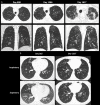
A, Mixed-phenotype chronic lung allograft dysfunction in a 33-year-old woman. CT images obtained on posttransplant day 808 show patchy subpleural consolidation and ground-glass opacities in the right upper lobe. CT images obtained on day 1286 show right upper lobe–predominant patchy ground-glass opacities and subpleural reticulation with volume loss. CT images obtained on day 1617 show progressive bilateral apical pleural thickening, subpleural fibrosis with traction bronchiectasis, volume loss, and architectural distortion, which are consistent with pleuroparenchymal fibroelastosis. B, CT images obtained on day 362 show right-middle-lobe airtrapping because of bronchial stenosis (not shown). CT images obtained on day 1557 show bilateral bronchiectasis and markedly worse bilateral airtrapping, evidenced by a lower-than-normal increase in attenuation and a lack of volume reduction, which are consistent with deteriorating accompanying obstruction. Additional patient information is found in Tables E1 and E2 (supplement).
Figure 8:
A, Mixed-phenotype chronic lung allograft dysfunction in a 33-year-old woman. CT images obtained on posttransplant day 808 show patchy subpleural consolidation and ground-glass opacities in the right upper lobe. CT images obtained on day 1286 show right upper lobe–predominant patchy ground-glass opacities and subpleural reticulation with volume loss. CT images obtained on day 1617 show progressive bilateral apical pleural thickening, subpleural fibrosis with traction bronchiectasis, volume loss, and architectural distortion, which are consistent with pleuroparenchymal fibroelastosis. B, CT images obtained on day 362 show right-middle-lobe airtrapping because of bronchial stenosis (not shown). CT images obtained on day 1557 show bilateral bronchiectasis and markedly worse bilateral airtrapping, evidenced by a lower-than-normal increase in attenuation and a lack of volume reduction, which are consistent with deteriorating accompanying obstruction. Additional patient information is found in Tables E1 and E2 (supplement).
Mixed-phenotype chronic lung allograft dysfunction in a 37-year-old man. CT studies obtained on posttransplant days 1817 and 2328 show worsening bilateral airtrapping manifested by a lower-than-anticipated decrease in volume and increased attenuation at expiration. CT images obtained on day 1817 show new focal peripheral consolidation in the posterior right upper lobe, which decreases in extent but fails to resolve on day 2328. Worsening volume loss and associated retraction of the oblique fissure is shown on images obtained on day 2328. Additional patient information is found in Tables E1 and E2 (supplement).
Figure 9:
Mixed-phenotype chronic lung allograft dysfunction in a 37-year-old man. CT studies obtained on posttransplant days 1817 and 2328 show worsening bilateral airtrapping manifested by a lower-than-anticipated decrease in volume and increased attenuation at expiration. CT images obtained on day 1817 show new focal peripheral consolidation in the posterior right upper lobe, which decreases in extent but fails to resolve on day 2328. Worsening volume loss and associated retraction of the oblique fissure is shown on images obtained on day 2328. Additional patient information is found in Tables E1 and E2 (supplement).
Hematoxylin-eosin–stained section from a lung at autopsy in a patient with chronic lung allograft dysfunction (imaging not available). A, Scanned micrograph shows typical features of acute fibrinous and organizing pneumonia (AFOP) associated with organizing alveolar fibroelastosis. The changes are distributed on both sides of an interlobular septum (ILS) in a perilobular pattern. Fibrinous pleuritis with edema has widened the pleura (pl). The horizontal bar is equivalent to 1.25 mm. B, Higher-magnification image of the region of AFOP shows compact fibrin balls occupying alveolar spaces (arrow) that can be seen separating away from the alveolar septa.
Figure 10:
Hematoxylin-eosin–stained section from a lung at autopsy in a patient with chronic lung allograft dysfunction (imaging not available). A, Scanned micrograph shows typical features of acute fibrinous and organizing pneumonia (AFOP) associated with organizing alveolar fibroelastosis. The changes are distributed on both sides of an interlobular septum (ILS) in a perilobular pattern. Fibrinous pleuritis with edema has widened the pleura (pl). The horizontal bar is equivalent to 1.25 mm. B, Higher-magnification image of the region of AFOP shows compact fibrin balls occupying alveolar spaces (arrow) that can be seen separating away from the alveolar septa.
Mixed-phenotype chronic lung allograft dysfunction in a 26-year-old woman. CT images obtained on posttransplant day 1447 demonstrate a multifocal bilateral upper lobe and peripherally predominant peribronchovascular ground-glass opacities and interlobular septal thickening (arrowheads), accompanied by elevated bronchoalveolar lavage and peripheral blood eosinophilia consistent with an eosinophilic pneumonia pattern, which resolves at follow-up CT on day 1611. CT images obtained on day 1651 demonstrate increased bronchial wall thickening and new multifocal peripheral consolidation with a reverse halo sign (arrow), in keeping with the organizing pneumonia pattern in the apicoposterior segment of the left upper lobe. Additional patient information is found in Tables E1 and E2 (supplement).
Figure 11:
Mixed-phenotype chronic lung allograft dysfunction in a 26-year-old woman. CT images obtained on posttransplant day 1447 demonstrate a multifocal bilateral upper lobe and peripherally predominant peribronchovascular ground-glass opacities and interlobular septal thickening (arrowheads), accompanied by elevated bronchoalveolar lavage and peripheral blood eosinophilia consistent with an eosinophilic pneumonia pattern, which resolves at follow-up CT on day 1611. CT images obtained on day 1651 demonstrate increased bronchial wall thickening and new multifocal peripheral consolidation with a reverse halo sign (arrow), in keeping with the organizing pneumonia pattern in the apicoposterior segment of the left upper lobe. Additional patient information is found in Tables E1 and E2 (supplement).
Mixed-phenotype chronic lung allograft dysfunction in a 68-year-old man. CT images obtained on posttransplant day 1096 demonstrate nodular subpleural consolidation in the apical right upper lobe (arrow). CT images obtained on day 1321 show new upper lobe–predominant perilobular ground-glass opacities and dense consolidation with associated bronchial dilatation and volume loss, as well as an organizing pneumonia reaction pattern with improved but persistent findings on images obtained on day 1350 (arrowheads). Additional patient information is found in Tables E1 and E2 (supplement).
Figure 12:
Mixed-phenotype chronic lung allograft dysfunction in a 68-year-old man. CT images obtained on posttransplant day 1096 demonstrate nodular subpleural consolidation in the apical right upper lobe (arrow). CT images obtained on day 1321 show new upper lobe–predominant perilobular ground-glass opacities and dense consolidation with associated bronchial dilatation and volume loss, as well as an organizing pneumonia reaction pattern with improved but persistent findings on images obtained on day 1350 (arrowheads). Additional patient information is found in Tables E1 and E2 (supplement).

References

    1. Chambers DC, Cherikh WS, Harhay MO, et al. The International Thoracic Organ Transplant Registry of the International Society for Heart and Lung Transplantation: thirty-sixth adult lung and heart-lung transplantation report-2019; focus theme: donor and recipient size match. J Heart Lung Transplant 2019;38(10):1042–1055. - PMC - PubMed
    1. Chambers DC, Yusen RD, Cherikh WS, et al. The Registry of the International Society for Heart and Lung Transplantation: thirty-fourth adult lung and heart-lung transplantation report-2017; focus theme: allograft ischemic time. J Heart Lung Transplant 2017;36(10):1047–1059. - PubMed
    1. Verleden GM, Glanville AR, Lease ED, et al. Chronic lung allograft dysfunction: definition, diagnostic criteria, and approaches to treatment: a consensus report from the Pulmonary Council of the ISHLT. J Heart Lung Transplant 2019;38(5):493–503. - PubMed
    1. Sumpter TL, Wilkes DS. Role of autoimmunity in organ allograft rejection: a focus on immunity to type V collagen in the pathogenesis of lung transplant rejection. Am J Physiol Lung Cell Mol Physiol 2004;286(6):L1129–L1139. - PubMed
    1. Glanville AR. Bronchoscopic monitoring after lung transplantation. Semin Respir Crit Care Med 2010;31(2):208–221. - PubMed

LinkOut - more resources