Case Reports
. 2021 Jan 28;3(1):e200469.
doi: 10.1148/ryct.2021200469. eCollection 2021 Feb.

Acute Myocarditis Associated with Acute Myocardial Infarction

Affiliations
Case Reports

Acute Myocarditis Associated with Acute Myocardial Infarction

Maycon Juglas Linhares Magalhães et al. Radiol Cardiothorac Imaging. .

Abstract

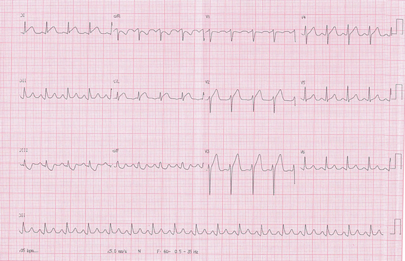
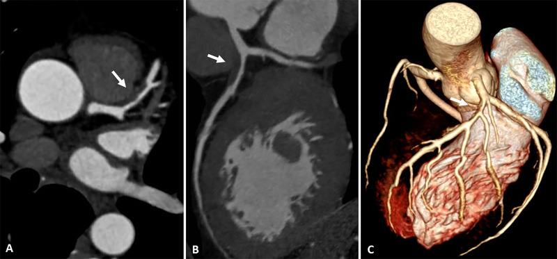
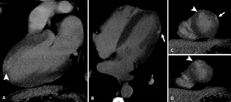
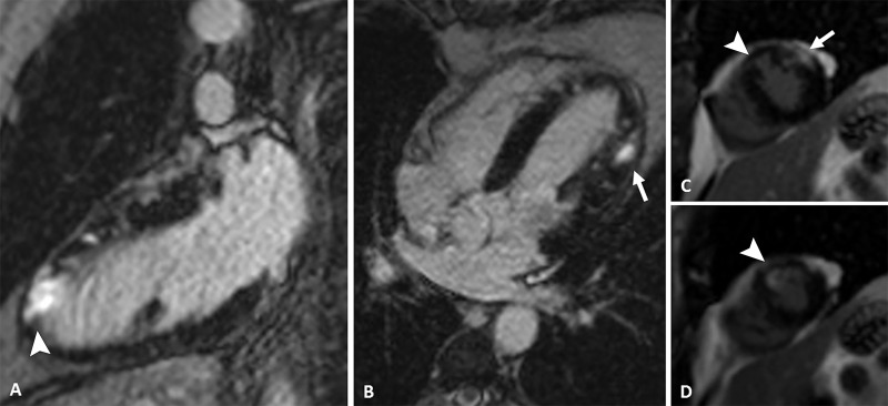
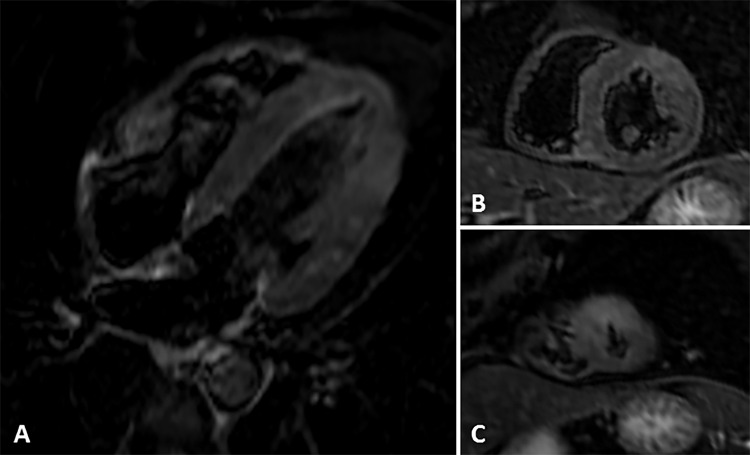
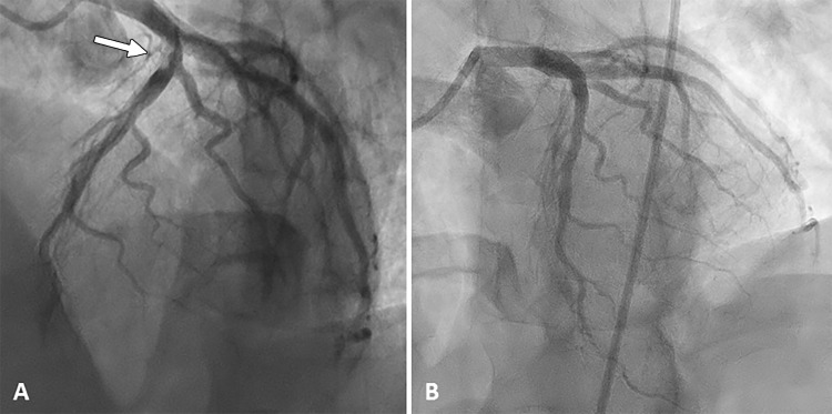
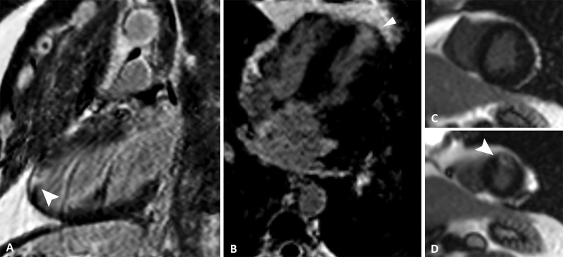
Concomitant acute myocarditis and acute coronary thrombosis is a rare presentation of acute chest pain in the emergency department, although the association between acute infections with a variety of pathogens and an increased risk of myocardial infarction has been reported. A case of acute myocardial infarction associated with acute myocarditis caused by coronavirus 229E in a middle-aged man without risk factors for coronary artery disease is described here. Coronary CT angiography with late enhancement protocol revealed areas of myocarditis and infarction, and cardiac MRI and coronary angiography were then performed. © RSNA, 2021.


Conflict of interest statement

Disclosures of Conflicts of Interest: M.J.L.M. disclosed no relevant relationships. L.d.P.G.d.F. disclosed no relevant relationships. L.P.S.B. disclosed no relevant relationships. M.C.S. disclosed no relevant relationships. P.O.G. disclosed no relevant relationships. A.C.d.A.B. disclosed no relevant relationships. P.G.M.d.B.e.S. Activities related to the present article: disclosed no relevant relationships. Activities not related to the present article: disclosed grants/grants pending to author’s institution from Bayer, Pfizer, and Roche Diagnostics. Other relationships: disclosed no relevant relationships. V.F. disclosed no relevant relationships.

Figures

Electrocardiogram reveals diffuse ST-segment elevation.
Figure 1:
Electrocardiogram reveals diffuse ST-segment elevation.
Images from coronary CT angiography. A, Axial, B, oblique multiplanar reconstruction, and, C, tridimensional reconstruction show an atherosclerotic plaque (arrow) in the proximal segment of anterior descending artery with severe luminal narrowing.
Figure 2:
Images from coronary CT angiography. A, Axial, B, oblique multiplanar reconstruction, and, C, tridimensional reconstruction show an atherosclerotic plaque (arrow) in the proximal segment of anterior descending artery with severe luminal narrowing.
Images from coronary CT angiography with late enhancement protocol. A, Two-chamber,B, four-chamber, and, C, D, short-axis views show late-enhancing mesoepicardial area (arrow) in the lateral apical segment suggestive of myocarditis and a subendocardial area (arrowhead) in the anterior apical segment suggestive of myocardial infarction.
Figure 3:
Images from coronary CT angiography with late enhancement protocol. A, Two-chamber, B, four-chamber, and, C, D, short-axis views show late-enhancing mesoepicardial area (arrow) in the lateral apical segment suggestive of myocarditis and a subendocardial area (arrowhead) in the anterior apical segment suggestive of myocardial infarction.
Images from phase-sensitive inversion-recovery cardiac MRI. A, Two-chamber, B, four-chamber, and, C, D, short-axis views show late-enhancing mesoepicardial area (arrow) in the lateral apical segment suggestive of myocarditis and a subendocardial area (arrowhead) in the anterior apical segment suggestive of myocardial infarction.
Figure 4:
Images from phase-sensitive inversion-recovery cardiac MRI. A, Two-chamber, B, four-chamber, and, C, D, short-axis views show late-enhancing mesoepicardial area (arrow) in the lateral apical segment suggestive of myocarditis and a subendocardial area (arrowhead) in the anterior apical segment suggestive of myocardial infarction.
Images from short τ inversion recovery cardiac MRI. A, Four-chamber and, B, C, short-axis views show edema in the mid anterolateral wall and lateral apical wall and in the anterior apical segment..
Figure 5:
Images from short τ inversion recovery cardiac MRI. A, Four-chamber and, B, C, short-axis views show edema in the mid anterolateral wall and lateral apical wall and in the anterior apical segment..
Images from coronary angiography. A, An ulcerated lesion causing 80% luminal narrowing in the proximal segment of the anterior descending artery, with a high thrombotic burden (arrow). B, After percutaneous coronary intervention with a drug-eluting stent.
Figure 6:
Images from coronary angiography. A, An ulcerated lesion causing 80% luminal narrowing in the proximal segment of the anterior descending artery, with a high thrombotic burden (arrow). B, After percutaneous coronary intervention with a drug-eluting stent.
Images from phase-sensitive inversion-recovery cardiac MRI. A, Two-chamber, B, four-chamber, and, C, D, short-axis views show late-enhancing subendocardial area (arrowhead) in the anterior apical segment suggestive of myocardial infarction. The late-enhancing mesoepicardial area in the lateral apical segment, compatible with myocarditis in the previous examination, was no longer seen.
Figure 7:
Images from phase-sensitive inversion-recovery cardiac MRI. A, Two-chamber, B, four-chamber, and, C, D, short-axis views show late-enhancing subendocardial area (arrowhead) in the anterior apical segment suggestive of myocardial infarction. The late-enhancing mesoepicardial area in the lateral apical segment, compatible with myocarditis in the previous examination, was no longer seen.

References

    1. Maron BJ, Towbin JA, Thiene G, et al. Contemporary definitions and classification of the cardiomyopathies: an American Heart Association Scientific Statement from the Council on Clinical Cardiology, Heart Failure and Transplantation Committee; Quality of Care and Outcomes Research and Functional Genomics and Translational Biology Interdisciplinary Working Groups; and Council on Epidemiology and Prevention. Circulation 2006;113(14):1807–1816. - PubMed
    1. Luetkens JA, Faron A, Isaak A, et al. Comparison of original and 2018 Lake Louise criteria for diagnosis of acute myocarditis: results of a validation cohort. Radiol Cardiothorac Imaging 2019;1(3):e190010. - PMC - PubMed
    1. Kindermann I, Kindermann M, Kandolf R, et al. Predictors of outcome in patients with suspected myocarditis. Circulation 2008;118(6):639–648 [Published correction appears in Circulation 2008;118(12): e493.]. - PubMed
    1. Baudry G, Bouleti C, Iung B, et al. Diagnosis of acute myocarditis with dual source cardiac tomography. Int J Cardiol 2015;179:256–257. - PubMed
    1. Kwong JC, Schwartz KL, Campitelli MA, et al. Acute Myocardial Infarction after Laboratory-Confirmed Influenza Infection. N Engl J Med 2018;378(4):345–353. - PubMed

Publication types