

doi: 10.1148/ryct.2021210008.
eCollection 2021 Feb.
Axillary Lymphadenopathy After mRNA COVID-19 Vaccination
Affiliations
- PMID: 33778667
- PMCID: PMC7861140
- DOI: 10.1148/ryct.2021210008
Item in Clipboard
Axillary Lymphadenopathy After mRNA COVID-19 Vaccination
Radiol Cardiothorac Imaging.
.
No abstract available
Conflict of interest statement
Dr. Richard Ahn owns 20 shares of Pfizer stock. Dr. Richard Ahn has no other relationship and no funding from Pfizer. Dr. Suhny Abbara reports royalty income from Elsevier/Amirsys. Dr. Cecelia Brewington reports grant funding from Cannon Medical Systems. Dr. Ann Mootz reports no conflicts of interest.
Figures
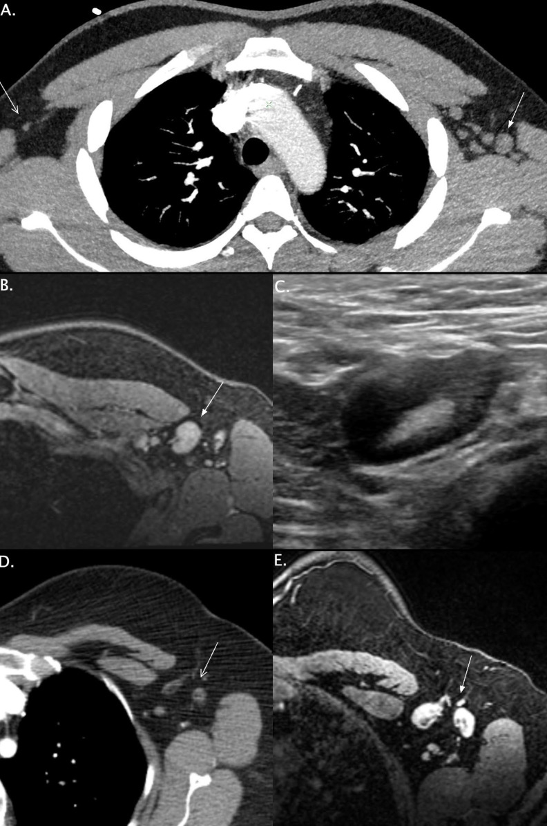
Axial CTA-MIP images demonstrating normal right axillary lymph nodes (A, open arrow) and unilateral left axillary lymphadenopathy (A, closed arrow) 7-days after administration of the second dose of an mRNA-COVID-19 vaccine in a 32-year-old man. The short axis of the lymph nodes measured up to 1.5 cm. Axial breast cancer screening MRI image (B, closed arrow) demonstrating a 2 cm left axillary lymph node seen 13 days after ipsilateral administration of the first dose of an mRNA COVID-19 vaccine in a 34-year-old female. Axillary ultrasound of the same patient (C) demonstrates a 2 cm likely reactive lymph node with diffuse cortical thickening. Follow-up imaging in 4-6 weeks was recommended. Axial CT image in a 39-year-old woman demonstrating normal left axillary lymph nodes (D, open arrow). Axial breast cancer screening MRI image ∼ 200 days later in the same patient (E, closed arrow) demonstrating new left axillary adenopathy measuring up to 1.4 cm in short axis 8 days after an mRNA COVID-19 vaccination. The patient was recalled for left axillary ultrasound. The involved lymph nodes in all three cases typically drain the arm (1). All patients were asymptomatic. Lymphadenopathy was reported in 64 patients receiving the BNT162b2-mRNA-COVID-19 vaccine compared to 6 in the placebo group (2). This potential association should be recognized in lung cancer screening, oncologic imaging and breast imaging as it is a differential consideration in workup of metastatic disease. While these cases are observational, further studies may investigate their association with a robust immune response.
References
-
- Ecanow JS, Abe H, Newstead GM, Ecanow DB, Jeske JM. Axillary Staging of Breast Cancer: What the Radiologist Should Know. Radiographics. 2013;33(6):1589-612. doi: 10.1148/rg.336125060. PubMed PMID: 24108553. - PubMed
-
- Pfizer-BioNTech COVID-19 Vaccine; FDA Briefing Document . Vaccines and Related Biological Products Advisory Committee Meeting. December, 10 2020
LinkOut - more resources
Full Text Sources
Other Literature Sources
