Review
. 2019 Nov 29;1(2):e190002.
doi: 10.1148/rycan.2019190002. eCollection 2019 Nov.

Renal Cell Carcinoma Ablation: Preprocedural, Intraprocedural, and Postprocedural Imaging

Affiliations
Review

Renal Cell Carcinoma Ablation: Preprocedural, Intraprocedural, and Postprocedural Imaging

Winston B Joe et al. Radiol Imaging Cancer. .

Abstract

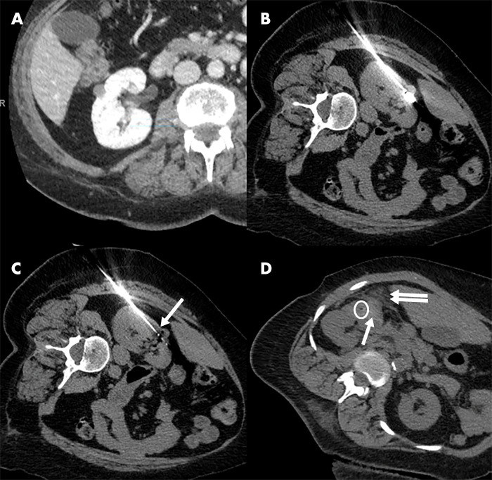
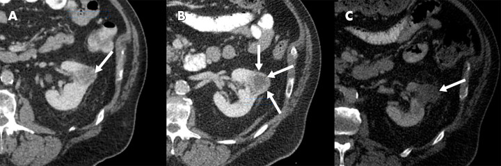
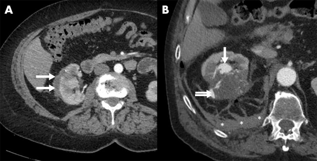
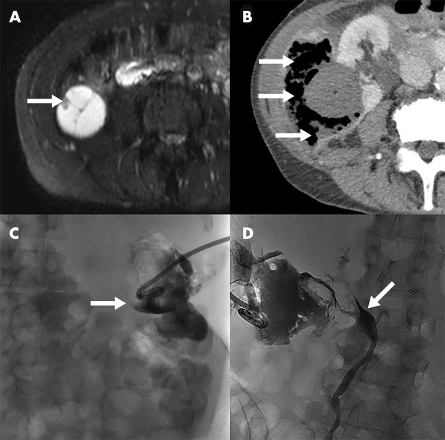
The rising incidence of renal cell carcinoma (RCC) in recent decades necessitates careful consideration of additional treatment options, especially for patients who may be poor surgical candidates. An emerging body of evidence suggests that ablation may be performed effectively and safely even in patients with multiple comorbidities. Accordingly, clinical guidelines now include thermal ablation as an alternative for such patients with localized tumors that are 4.0 cm or smaller. Recent experience with these minimally invasive techniques has led to a greater understanding of the imaging findings that merit close attention when ablation is anticipated, or after it is performed. These imaging findings may guide the interventionalist's perception of the risks, technical challenges, and likelihood of treatment success associated with RCC ablation. The present review provides an overview of clinically relevant radiologic findings during the preprocedural, intraprocedural, and postprocedural period in the context of image-guided renal ablation. Keywords: Interventional-Body, Kidney, Percutaneous, Urinary © RSNA, 2019.


Conflict of interest statement

Disclosures of Conflicts of Interest: W.B.J. disclosed no relevant relationships. J.G.Z. disclosed no relevant relationships. A.J.G. Activities related to the present article: disclosed no relevant relationships. Activities not related to the present article: Serves as a consultant for BTG and AngioDynamics.

Figures

Figure 1:
Figure 1:
US appearance of renal cell carcinoma in a 61-year-old woman. A, Gray-scale US image shows a hyperechoic, well-circumcised, 3.5-cm mass at the lower pole of the right kidney. B, Doppler US image demonstrates evidence of vascularization within the mass; C, contrast-enhanced US confirms tumor vascularization; D, real-time gray-scale US image immediately after microwave ablation shows areas of gas with possible residual echogenicity; E, real-time contrast-enhanced US image shows no residual enhancing material in the ablation bed, confirming treatment effect.
Figure 2:
Figure 2:
CT appearance of renal cell carcinoma in an 85-year-old man. A, Axial unenhanced CT image shows exophytic mass in the left lateral kidney measuring 13 HU; B, axial CT image in the corticomedullary phase demonstrates substantial mass enhancement (63 HU); C, axial CT image in the nephrographic phase with continued enhancement (62 HU); D, axial dual-energy CT virtual unenhanced image with mass measuring 15 HU; E, axial dual-energy CT iodine density image shows the quantitative iodine content within the mass to be 4.2 mg/mL; F, coronal CT images showing mass in the nephrographic phase.
Figure 3:
Figure 3:
MRI appearance of renal cell carcinoma in a 59-year-old man. A, Axial T2-weighted MR image shows exophytic 3.7-cm left renal mass; B, unenhanced axial T1-weighted MR image shows isointense left renal mass with punctate foci of hyperintensity (white arrows); C, axial contrast-enhanced T1-weighted MR image in arterial phase shows tumor enhancement; D, contrast-enhanced axial T1-weighted MR image in venous phase shows persistent enhancement; E, subtracted arterial phase MR image confirms enhancement within the mass; F, axial subtracted portal venous phase MR image confirms punctate foci of hyperintensity seen on T1-weighted MRI are nonenhancing (black arrows); G, axial diffusion-weighted image shows the left renal mass to be hyperintense to renal parenchyma; H, corresponding axial apparent diffusion coefficient map shows relatively hypointense mass, confirming diffusion restriction.
Figure 4:
Figure 4:
Percutaneous cryoablation of renal cell carcinoma and common postprocedural findings in an 89-year-old woman. A, Preprocedural axial contrast-enhanced CT image shows exophytic 3.4-cm mass in lower pole of right kidney (white arrow); B, axial unenhanced CT image during procedural planning shows patient in prone position with marking grid overlying mass (black arrows); C, intraprocedural axial CT image shows one of four cryoablation probes within the mass prior to first freeze-thaw cycle; D, intraprocedural axial CT image following second freeze-thaw cycle shows ice ball (arrowheads) with complete tumor coverage and appropriate margins without bowel encroachment (bracket); E, axial CT image obtained immediately after removal of probes showing common postprocedural findings including: gas in region of removed probe (white arrow) which may also appear as a hyperdense line, a visible ice ball (black arrow), and hyperdense areas representing blood products (arrowheads); F, subtracted MR image in portal venous phase at 1-year follow-up shows no residual enhancement in the ablation bed (white arrow) indicating complete response.
Figure 5:
Figure 5:
Percutaneous microwave ablation (MWA) of renal cell carcinoma and common intraprocedural findings. A, Preprocedural axial contrast-enhanced CT image in portal venous phase shows an exophytic 1.5-cm right renal mass in a 60-year-old woman, less than 1 cm from the colon; B, intraprocedural axial unenhanced CT image shows MWA probe in place; C, intraprocedural axial unenhanced CT image demonstrates gas bubbles (arrow) within the ablation zone, a common finding during MWA; D, other common findings after MWA demonstrated on axial unenhanced CT image in a separate 61-year-old woman including: gas (circled), hyperdensity (arrow) representing blood products, and a small amount of asymptomatic perinephric hematoma (double arrows).
Figure 6:
Figure 6:
Clinically relevant findings at follow-up. A, Axial contrast-enhanced CT image in portal venous phase in a 65-year-old man demonstrates an exophytic 2.6-cm left renal mass (arrow) subsequently treated with cryoablation; B, axial unenhanced CT image 6 months after ablation shows the ablation zone with a benign fibrofatty rim (white arrows); C, axial contrast-enhanced CT image at the same visit demonstrates no enhancement of the ablation zone (white arrow), consistent with a complete treatment response.
Figure 7:
Figure 7:
Clinically relevant findings at follow-up. A, Axial contrast-enhanced CT image in portal venous phase in a 72-year-old man demonstrates a 3.4-cm left renal mass (white arrow) subsequently treated with microwave ablation (MWA); B, axial contrast-enhanced CT image at 6-month follow-up shows residual tumor with nodular enhancement (arrows); C, axial contrast-enhanced CT image 3 months after repeat MWA shows no remaining enhancement in the ablation bed (white arrow).
Figure 8:
Figure 8:
Hemorrhage after percutaneous ablation; A, Axial contrast-enhanced CT image in portal venous phase obtained 3 months after microwave ablation of a 1.5-cm right renal mass demonstrates a small perinephric hematoma (arrows) without extravasation of contrast material. The hematoma was clinically insignificant as the patient was asymptomatic and without any significant changes to laboratory values. B, Axial contrast-enhanced CT in arterial phase obtained 1 day after cryoablation of a 6.5-cm right renal mass due to an episode of hematuria and hypotension shows a pseudoaneurysm (arrow) with extravasation of contrast material (double arrow) leading to a large peri-nephric hematoma (*). This was successfully treated using coil embolization and the patient was discharged the following day. Imaging obtained at 3 months (not shown) demonstrated a complete response to treatment without any additional complications.
Figure 9:
Figure 9:
Abscess formation after percutaneous ablation; A, Axial T2-weighted MR image obtained in a 54-year-old woman demonstrates a complex cystic mass with a nodule (white arrow) that was subsequently treated with cryoablation using hydrodissection to move the colon away from the ablation zone; B, the patient presented 3 weeks later with abdominal pain and fever. An axial contrast-enhanced CT image demonstrated a perinephric abscess (arrows). The abscess was treated with percutaneous drainage. C, Radiograph obtained during percutaneous abscess drainage catheter placement shows contrast material flowing into the colon (arrow), confirming a fistula with the bowel. D, Radiograph obtained during percutaneous abscess drainage catheter placement shows contrast material in the renal collecting system (arrow), confirming a fistula. This was treated with ureteric stent placement. A small perforation of the bowel was noted at colonoscopy which had healed at repeat colonoscopy 3 months later. Percutaneous drainage catheter and ureteral stents then were removed, and the patient experienced no long-term sequelae.

Similar articles

Cited by

References

    1. Hollingsworth JM, Miller DC, Daignault S, Hollenbeck BK. Rising incidence of small renal masses: a need to reassess treatment effect. J Natl Cancer Inst 2006;98(18):1331–1334. - PubMed
    1. Siegel RL, Miller KD, Jemal A. Cancer statistics, 2019. CA Cancer J Clin 2019;69(1):7–34. - PubMed
    1. Motzer RJ, Jonasch E, Agarwal N, et al. . NCCN Clinical Practice Guidelines in Oncology: Kidney Cancer. National Comprehensive Cancer Network. https://www.nccn.org/professionals/physician_gls/pdf/kidney.pdf. Published February 6, 2019. Accessed February 10, 2019. - PubMed
    1. Campbell S, Uzzo RG, Allaf ME, et al. . Renal mass and localized renal cancer: AUA guideline. J Urol 2017;198(3):520–529. - PubMed
    1. Rivero JR, De La Cerda J 3rd, Wang H, et al. . Partial nephrectomy versus thermal ablation for clinical stage T1 renal masses: systematic review and meta-analysis of more than 3,900 patients. J Vasc Interv Radiol 2018;29(1):18–29. - PubMed

MeSH terms